Comparative genomics of the major parasitic worms
Abstract
Parasitic
nematodes (roundworms) and platyhelminths (flatworms) cause
debilitating chronic infections of humans and animals, decimate crop
production and are a major impediment to socioeconomic development.
Here
we report a broad comparative study of 81 genomes of parasitic and
non-parasitic worms. We have identified gene family births and hundreds
of expanded gene families at key nodes in the phylogeny that are
relevant to parasitism. Examples include gene families that modulate
host immune responses, enable parasite migration though host tissues or
allow the parasite to feed. We reveal extensive lineage-specific
differences in core metabolism and protein families historically
targeted for drug development. From an in silico screen, we have
identified and prioritized new potential drug targets and compounds for
testing. This comparative genomics resource provides a much-needed boost
for the research community to understand and combat parasitic worms.
Main
Over a quarter of humans are infected with parasitic nematodes (roundworms) or platyhelminths (flatworms)
1.
Although rarely lethal, infections are typically chronic, leading to
pain, malnutrition, physical disabilities, delayed development,
deformity, social stigma or a burden on family members caring for the
afflicted. These diseases encompass many of the most neglected tropical
diseases and attract little research investment. Parasitic nematodes and
platyhelminths impede economic development through human disability,
and billions of dollars of lost production in the livestock
2 and crop
3 industries.
Few
drugs are available to treat worm infections. Repeated mass
administration of monotherapies is increasing the risk of resistance to
human anthelmintics
4 and has driven widespread resistance in farm animals
5. There are no vaccines for humans, and few for animals
6. The commonly used nematicides of plant parasites are environmentally toxic
7, and need replacement.
The
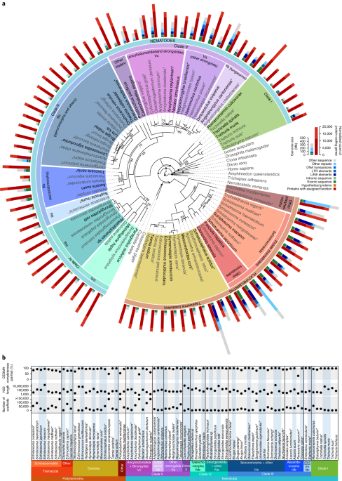
phylum Nematoda is part of the superphylum Ecdysozoa and has five major
clades (I to V), four of which contain human-infective parasites and
are analyzed here (Fig. 1).
The phylum Platyhelminthes is part of the superphylum Lophotrochozoa
and the majority of parasite species are cestodes (tapeworms) and
trematodes (flukes). Comparing the genomes of parasites from these two
phyla may reveal common strategies employed to subvert host defenses and
drive disease processes.
We have combined 36 published genomes
8,9,10,11,12,13,14,15,16,17,18,19,20,21,22,23,24,25,26,27,28,29,30,31,32,33,34
with new assemblies for 31 nematode and 14 platyhelminth species into a
large genome comparison of parasitic and non-parasitic worms. We have
used these data to identify gene families and processes associated with
the major parasitic groups. To accelerate the search for new
interventions, we have mined the dataset of more than 1.4 million genes
to predict new drug targets and drugs.
Results
Genomic diversity in parasitic nematodes and platyhelminths
We
have produced draft genomes for 45 nematode and platyhelminth species
and predicted 0.8 million protein-coding genes, with 9,132–17,274 genes
per species (5–95% percentile range; see
Methods, Supplementary Tables
1–
3, Supplementary Fig.
1 and Supplementary Notes
1.1 and
1.2). We combined these new data with 36 published worm genomes—comprising 31 parasitic
8,9,10,11,12,13,14,15,16,17,18,19,20,21,22,23,24,25,26,27,28,29,30 and five free-living
18,31,32,33,34 species—and 10 outgroups
35,36,37,38,39,40,41,42,43,44 from other animal phyla, into a comparative genomics resource of 91 species (Fig.
1 and Supplementary Tables
2 and
4).
There was relatively little variation in gene set completeness
(coefficient of variation, c.v. = 0.15) among the nematodes and
platyhelminths, despite variation in assembly contiguity (c.v. = 8.5;
Fig.
1b and Supplementary Table
2). Nevertheless, findings made using a subset of high-quality assemblies that were designated ‘tier 1’ (
Methods and Supplementary Table
4) were corroborated against all species.
Genome
size varied greatly within each phylum, from 42 to 700 Mb in nematodes,
and from 104 to 1,259 Mb in platyhelminths. In a small number of cases,
size estimates may have been artifactually inflated by high
heterozygosity causing alternative haplotypes to be represented within
the assemblies (Supplementary Note
1.3 and Supplementary Table
2a).
A more important factor appeared to be repeat content that ranged
widely, from 3.8 to 54.5% (5–95% percentile; Supplementary Table
5).
A multiple regression model, built to rank the major factors driving
genome size variation, identified long terminal repeat transposons,
simple repeats, assembly quality, DNA transposable elements, total
length of introns and low complexity sequence as being the most
important (Supplementary Note
1.3,
Methods and Supplementary Table
6). Genome size variation is thus largely due to non-coding elements, as expected
45,
including repetitive and non-repetitive DNA, suggesting it is either
non-adaptive or responding to selection only at the level of overall
genome size.
Gene family births and expansions
We inferred gene families from the predicted proteomes of the 91 species using Ensembl Compara
46. Of the 1.6 million proteins, 1.4 million were placed into 108,351 families (Supplementary Note
2.1 and
Supplementary Data), for which phylogenetic trees were built and orthology and paralogy inferred (
Methods, Supplementary Fig.
2 and Supplementary Table
7). Species trees inferred from 202 single-copy gene families that were present in at least 25% of species (Fig.
1),
or from presence/absence of gene families, largely agreed with the
expected species and clade relationships, except for a couple of known
contentious issues (Supplementary Fig.
3, Supplementary Note
2.2 and
Methods).
The
species in our dataset contained significant novelty in gene content.
For example, ~28,000 parasitic nematode gene families contained members
from two or more parasitic species but were absent from
Caenorhabditis elegans and 47% of gene families lacked any functional annotation (Supplementary Note
2.1 and
Methods). The latter families tended to be smaller than those with annotations (Supplementary Fig.
4)
and, in many cases, correspond to families that are so highly diverged
that ancestry cannot be traced, reflecting the huge breadth of
unexplored parasite biology.
Gene families specific to particular
parasite clades are likely to reflect important aspects of parasite
biology and possible targets for new antiparasitic interventions. At key
nodes in the phylogeny that are relevant to parasitism, we identified
5,881 families with apparent clade-specificity (synapomorphies;
Supplementary Note
2.3,
Methods and Supplementary Table
8),
although our ability to discriminate truly parasite-specific clades was
limited by the low number of free-living species. The apparent
synapomorphies were either gene family births, or subfamilies that were
so diverged from their homologues that they appeared as separate
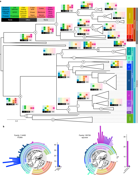
families. Functional annotation of these families was diverse (Fig.
2),
but they were frequently associated with sensory perception (such as
G-protein coupled receptors; GPCRs), parasite surfaces (platyhelminth
tegument or nematode cuticle maintenance proteins) and protein
degradation (proteases and protease inhibitors).
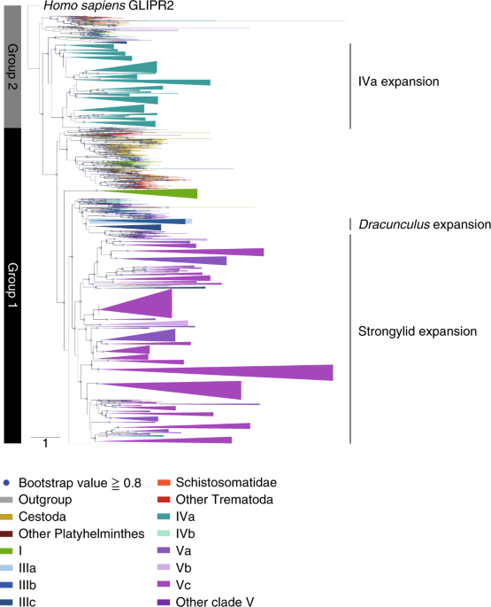
Among nematodes, clade IVa (which includes
Strongyloides spp.; Fig.
1)
showed the highest number of clade-specific families, including a novel
ferrochelatase-like family. Most nematodes lack functional
ferrochelatases for the last step of haem biosynthesis
47,
but harbor ferrochelatase-like genes of unknown function, to which the
synapomorphic clade IVa family was similar (Supplementary Fig.
5 and
Methods).
Exceptions are animal parasites in nematode clades III (for example
ascarids and filaria) and IV that acquired a functional ferrochelatase
via horizontal gene transfer
48,49.
Within the parasitic platyhelminths, a clade-specific
inositol-pentakisphosphate 2-kinase (IP2K) was identified. In some
species of
Echinococcus tapeworms, IP2K produces inositol
hexakisphosphate nanodeposits in the extracellular wall (the laminated
layer) that protects larval metacestodes
50. The deposits increase the surface area for adsorption of host proteins and may promote interactions with the host
51.
Paralogous
expansions of gene families, particularly those that are large or
repeatedly involve related processes, can be evidence of adaptive
evolution. We searched among our 10,986 highest-confidence gene families
(those containing ≥10 genes from tier 1 species) for those that had
expanded in parasite clades. A combination of scoring metrics (
Methods)
reduced the list to 995 differentially distributed families with a bias
in copy number in at least one parasite clade. Twenty-five expansions
have previously been observed, including 21 with possible roles in
parasitism (Supplementary Fig.
6).
A further 43 were placed into major functional classes that
historically have been favored as drug targets (kinases, GPCRs, ion
channels and proteases
52; Supplementary Table
9a).
By
manually inspecting the distribution of the remaining 927 families
across the full species tree, we identified 176 families with striking
expansions (Supplementary Table
9a and Supplementary Note
2.4).
Thirty two had no functional annotation; for example, family 393312 was
highly expanded in clade Va nematodes (Supplementary Fig.
7 and Supplementary Table
9a).
Even when families could be functionally annotated to some extent (for
example, based on a protein domain), discerning their precise biological
role was a challenge. For example, a sulfotransferase family that was
expanded in flukes compared with tapeworms includes the
Schistosoma mansoni locus that is implicated in resistance to the drug oxamniquine
53 but the endogenous substrate for this enzyme is unknown (Supplementary Fig.
7j).
Among
the newly identified expansions, we focused on those with richer
functional information, especially where they were related to similar
biological processes. For instance, we identified several expansions of
gene families involved in innate immunity of the parasites, as well as
their development. These included families implicated in protection
against bacterial or fungal infections in nematode clade IVa (
bus-4 GT31 galactosyltransferase
54,
irg-355) and clades Va/Vc (lysozyme
56 and the dual oxidase
bli-357) (Supplementary Fig.
8a–d). In nematode clade IIIb, a family was expanded that contains orthologs of the
Parascaris coiled-coiled protein PUMA, involved in kinetochore biology
58 (Fig.
2b).
This expansion possibly relates to the evolution of chromatin
diminution in this clade, which results in an increased number of
chromosomes requiring correct segregation during metaphase
59. In nematode clade IVa and in
Bursaphelenchus, an expansion of a steroid kinase family (Supplementary Fig.
8e)
is suggestive of novelty in steroid-regulated processes in this group,
such as the switch between free-living or parasitic stages in
Strongyloides60.
Infections
with parasitic worms are typified by their chronicity and a plausible
involvement in host–parasite interactions is a recurring theme for many
of the families.
Taenia tapeworms and clade V strongylid nematodes (that is Va, Vb and Vc; Fig.
1)
contained two expanded families with apyrase domains that may have a
role in hydrolyzing ATP (a host danger signal) from damaged host tissue
61 (Fig.
2b and Supplementary Fig.
9a).
Moreover, many of the strongylid members also contained amine
oxidoreductase domains, possibly to reduce production of
pro-inflammatory amines, such as histamine, from host tissues
62.
In platyhelminths, we observed expansions of tetraspanin families that
are likely components of the host/pathogen interface. Described examples
show tetraspanins being part of extracellular vesicles released by
helminths within hosts
63; or binding the Fc domain of host antibodies
64; or being highly immunogenic
65 (Supplementary Fig.
9b,c).
In strongylids, especially clade Vc, an expansion of the fatty acid and
retinol-binding (FAR) family, implicated in host–parasite interaction
of plant- and animal-parasitic nematodes
66,67 (Supplementary Fig.
9d),
suggests a role in immune modulation. Repertoires of glycosyl
transferases have expanded in nematode clades Vc and IV, and tapeworms
(Supplementary Fig.
10a–c),
and may be used to evade or divert host immunity by modifying parasite
surface molecules directly exposed to the immune system
68; alternatively, surface glycoproteins may interact with lectin receptors on innate immune cells in an inhibitory manner
69.
An expanded chondroitin hydrolase family in nematode clade Vc may
possibly be used either for larval migration through host connective
tissue or to digest host intestinal walls (Supplementary Fig.
9e). Similarly, an expanded GH5 glycosyl hydrolase family contained schistosome members with egg-enriched expression
8,70 that may be used for traversing host tissues such as bladder or intestinal walls (Supplementary Fig.
9f).
In nematode clade I, we found an expansion of a family with the
PAN/Apple domain, which is implicated in attachment of some protozoan
parasites to host cells
71, and possibly modulates host lectin-based immune activation (Supplementary Fig.
9g).
The
SCP/TAPS (sperm-coating protein/Tpx/antigen 5/pathogenesis-related
protein 1) genes have been associated with parasitism through their
abundance, secretion and evidence of their role in immunomodulation
72
but are poorly understood. This diverse superfamily appeared as eight
expanded Compara families. A more comprehensive phylogenetic analysis of
the full repertoire of 3,167 SCP/TAPS sequences (Supplementary Note
2.5, Supplementary Table
10 and
Methods) revealed intra- and interspecific expansions and diversification over different evolutionary timescales (Fig.
3 and Supplementary Figs.
11a,b and
12).
In particular, the SCP/TAPS superfamily has expanded independently in
nematode clade V (18–381 copies in each species) and in clade IVa
parasites (39–166 copies) (Fig.
3 and Supplementary Fig.
11c).
Dracunculus medinensis
(Guinea worm) was unusual in being the only member of clade III to
display an expansion (66 copies), which may reflect modulation of the
host immune response during the tissue migration phase of its large
adult females.
Proteins historically targeted for drug development
Proteases, GPCRs, ion channels and kinases dominate the list of targets for existing drugs for human diseases
52,
and are attractive leads for developing new ones. We therefore explored
the diversity of these superfamilies across the nematodes and
platyhelminths (Supplementary Fig.
13, Supplementary Note
3 and
Methods).
Proteases
and protease inhibitors perform diverse functions in parasites,
including immunomodulation, host tissue penetration, modification of the
host environment (for example, anticoagulation) and digestion of blood
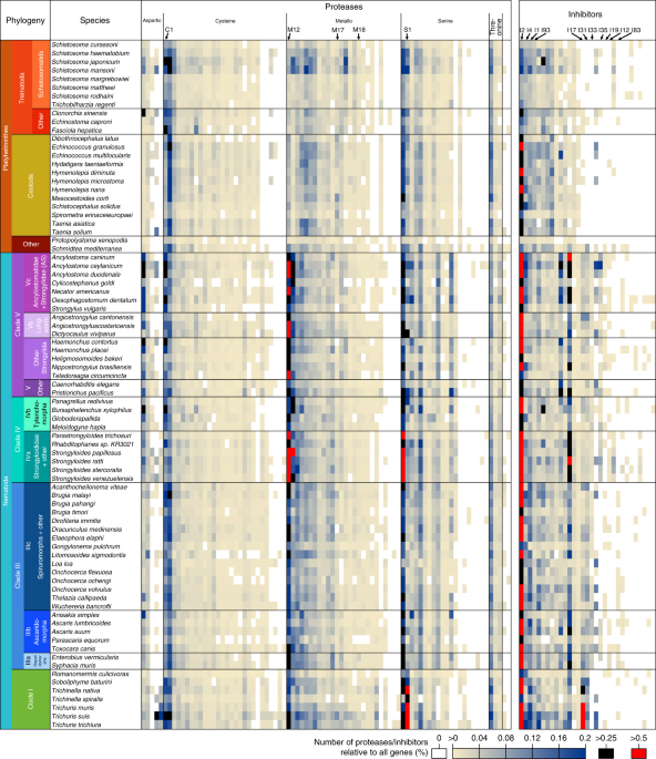
73. M12 astacins have particularly expanded in nematode clade IVa (five families), as previously reported
18, but there are two additional expansions in clades Vc and Vb (Fig.
4, Supplementary Fig.
14 and Supplementary Table
11). Because many of these species invade through skin (IVa, Vc; Supplementary Table
12) and migrate through the digestive system and lung (IVa, Vc, Vb; Supplementary Table
13),
these expansions are consistent with evidence that astacins are
involved in skin penetration and migration through connective tissue
74.
The cathepsin B C1-cysteine proteases are particularly expanded in
species that feed on blood (two expansions in nematode clades Vc and Va
30, with highest platyhelminth gene counts in schistosomatids and
Fasciola12; Supplementary Fig.
14). Indeed, they are involved in blood digestion in adult nematodes
75 and platyhelminths
76, but some likely have different roles such as larval development
77 and host invasion
78.
Different
protease inhibitors may modulate activity of parasite proteases or
protect parasitic nematodes and platyhelminths from degradation by host
proteases, facilitate feeding or manipulate the host response to the
parasite
79.
The I2 (Kunitz-BPTI) trypsin inhibitors are the most abundant protease
inhibitors across parasitic nematodes and platyhelminths (Fig.
4). An expansion of the I17 family, which includes secretory leukocyte peptidase inhibitor, was reported previously in
Trichuris muris17 but the striking confinement of this expansion to most of the parasites of clade I is now apparent (Fig.
4).
We also observed a notable family of α-2-macroglobulin (I39) protease
inhibitors that are present in all platyhelminths but expanded in
tapeworms (Supplementary Fig.
14).
The tapeworm α-2-macroglobulins may be involved in reducing blood
clotting at attachment or feeding sites; alternatively, they may
modulate the host immune response, since α-2-macroglobulins bind several
cytokines and hormones
80.
Chymotrypsin/elastase inhibitors (family I8) were particularly expanded
in clades Vc and IVa (consistent with upregulation of I8 genes in
Strongyloides parasitic stages
18) and to a lesser extent in clade IIIb (Fig.
4), consistent with evidence that they may protect
Ascaris from host proteases
81.
We also identified protein domain combinations that were specific to
either nematodes or platyhelminths (131 and 50 domain combinations,
respectively). Many of these involved protease and protease inhibitor
domains. In nematodes, several combinations included Kunitz protease
inhibitor domains, and in platyhelminths metalloprotease families M18
and M28 were found in novel combinations (Supplementary Table
14, Supplementary Note
3.2 and
Methods).
Of the 230 gene families annotated as GPCRs (Supplementary Figs.
13 and
15 and Supplementary Note
3.3),
only 21 were conserved across phyla. Chemosensory GPCRs, while abundant
in nematodes, were not identified in platyhelminths, although they are
identifiable in other Lophotrochozoa (such as Mollusca
82), suggesting that either the platyhelminths have lost this class or they are very divergent (Supplementary Table
15).
GPCR families lacking sequence similarity with known receptors included
the platyhelminth-specific rhodopsin-like orphan families (PROFs),
which are likely to be class A receptors and peptide responsive, and
several other fluke-specific non-PROF GPCR families. The massive
radiation of chemoreceptors in
C. elegans was unmatched in any
other nematode (87% versus ≤48% of GPCRs). All parasitic nematodes
possessed chemoreceptors, with the most in clade IVa, including several
large families synapomorphic to this clade (Supplementary Fig.
15), perhaps related to their unusual life cycles that alternate between free-living and parasitic forms.
Independent
expansion and functional divergence has differentiated the nematode and
platyhelminth pentameric ligand gated ion channels (Supplementary Fig.
16, Supplementary Table
16 and Supplementary Note
3.4). For example, glutamate signaling arose independently in platyhelminths and nematodes
83, and in trematodes the normal role of acetylcholine has been reversed, from activating to inhibitory
84.
Our analysis suggested the platyhelminth acetylcholine-gated anion
channels are most related to the Acr-26/27 group of nematode nicotinic
acetylcholine receptors that are the target of the anthelmintics
morantel and pyrantel
85, rather than to nematode acetylcholine-gated cation channels, targeted by nicotine and levamisole (Supplementary Fig.
17).
ABC transporters (Supplementary Table
17 and Supplementary Note
3.5) and kinases (Supplementary Note
3.6 and Supplementary Fig.
18)
showed losses and independent expansion within nematodes and
platyhelminths. The P-glycoprotein class of transporters, responsible
for the transport of environmental toxins and linked with anthelmintic
resistance, is expanded relative to vertebrates
86, with increased numbers in nematodes (Supplementary Fig.
19).
Metabolic reconstructions of nematodes and platyhelminths
In
the context of drug discovery, understanding the metabolic capabilities
of parasitic worms may reveal vulnerabilities that can be exploited in
target-based screens for new compounds. For each of the 81 nematode and
platyhelminth species, metabolism was reconstructed based on high
confidence assignment of enzyme classes (Supplementary Table
18a). The nematodes had a greater range of annotated enzymes per species than the platyhelminths (Supplementary Fig.
20a),
in part reflecting the paucity of biochemical studies in
platyhelminths. Because variation in assembly quality or divergence from
model organisms
87
could bias enzyme predictions, we identified losses of pathways and
differences in pathway coverage across different clades (Supplementary
Note
4,
Methods, Fig.
5 and Supplementary Fig.
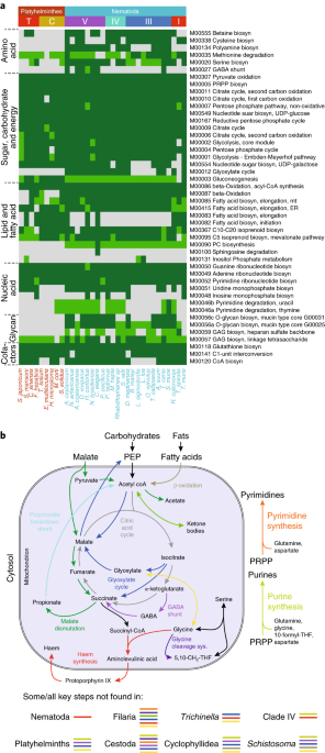
21). Pathways related to almost all metabolic superpathways in the Kyoto Encyclopedia of Genes and Genomes (KEGG)
88
showed significantly lower coverage for platyhelminths (versus
nematodes) and filaria (versus other nematodes) (Supplementary Fig.
20b).
In
contrast to most animals, nematodes possess the glyoxylate cycle that
enables conversion of lipids to carbohydrates, to be used for
biosyntheses (for example, during early development) and to avert
starvation
89. The glyoxylate cycle appears to have been lost independently in the filaria and
Trichinella species (Fig.
5a; M00012), both of which are tissue-dwelling obligate parasites. The filaria and
Trichinella have also independently lost alanine-glyoxylate transaminase that converts glyoxylate to glycine (Fig.
5b).
Glycine can be converted by the glycine cleavage system (GCS) to
5,10-methylenetetrahydrofolate, a useful one-carbon pool for
biosyntheses, and two key GCS proteins appear to have been lost
independently from filaria and tapeworms, suggesting their GCS is
non-functional (Supplementary Table
19e).
In addition, filaria have lost the ability to produce and use ketone
bodies, a temporary store of acetyl coenzyme A (CoA) under starvation
conditions (Supplementary Table
19b). The filaria lost these features after they diverged from
D. medinensis,
an outgroup to the filaria in clade IIIc that has a major difference in
its life cycle, namely, a free-living larval stage (Supplementary Table
12).
The
absence of multiple initial steps of pyrimidine synthesis was observed
in some nematodes, including all filaria (as previously reported
23) and tapeworms, suggesting they obtain pyrimidines from
Wolbachia endosymbionts or from their hosts, respectively (Supplementary Table
19f).
Similarly, all platyhelminths and some nematodes (especially clade IVa
and filaria IIIc) appear to lack key enzymes for purine synthesis
(Supplementary Table
19g) and rely on salvage instead. However, despite the widespread belief that nematodes cannot synthesize purines
90,91,
complete or near-complete purine synthesis pathways were found in most
members of clades I, IIIb and V. Nematodes are known to be unable to
synthesize haem
47 but the pathway was found in platyhelminths, including
S. mansoni (despite conflicting biochemical data
47) (Supplementary Table
19h and Supplementary Table
20i).
Genes
from the β-oxidation pathway, used to break down lipids as an energy
source, were not detected in schistosomes and some cyclophyllidean
tapeworms (
Hymenolepis,
Echinococcus; Fig.
5a, M00087; Supplementary Table
19a).
These species live in glucose-rich environments and may have evolved to
use glucose and glycogen as principal energy sources. However,
biochemical data suggest they do perform β-oxidation
92, so they may have highly diverged but functional β-oxidation genes.
The
lactate dehydrogenase (LDH) pathway is a major source of ATP in
anaerobic but glucose-rich environments. Platyhelminths have high
numbers of
LDH genes, as do blood-feeding
Ancylostoma hookworms (Supplementary Fig.
22g). Nematode clades Vc (including
Ancylostoma)
and IIIb have expansions of α-glucosidases that may break down starch
and disaccharides in host food to glucose (Supplementary Fig.
22a). Many nematodes and flatworms use malate dismutation as an alternative pathway for anaerobic ATP production
93.
The importance of the pathway for clade IIIb nematodes was reflected in
expanded families encoding two key pathway enzymes PEPCK and
methylmalonyl CoA epimerase, and the intracellular trafficking chaperone
for cobalamin (vitamin B-12), a cofactor for the pathway (Supplementary
Fig.
22c–e and Supplementary Table
9a).
A second cobalamin-related family (CobQ/CbiP) is clade IIIb-specific
and appears to have been gained by horizontal gene transfer from
bacteria (Supplementary Fig.
23a, Supplementary Note
2.6 and
Methods). A glutamate dehydrogenase family expanded in clade IIIb (Supplementary Fig.
22h)
is consistent with a GABA (γ-aminobutyric acid) shunt that helps
maintain redox balance during malate dismutation. In clade Va, an
expansion in the propionate breakdown pathway
94 (Supplementary Fig.
22f), suggested degradation of propionate, originating from malate dismutation or fermentation in the host’s stomach
95. Clade I nematodes have an acetate/succinate transporter that appeared to have been gained from bacteria (Supplementary Note
2.6 and
Methods), and may participate in acetate/succinate uptake or efflux (Supplementary Fig.
23b).
Identifying new anthelmintic drug targets and drugs
As
an alternative to a purely target-based approach that would require
extensive compound screening, we explored drug repurposing
possibilities. We developed a pipeline to identify the most promising
targets from parasitic nematodes and platyhelminths. These sequences
were used in searches of the ChEMBL database that contains curated
activity data on defined targets in other species and their associated
drugs and compounds (Supplementary Note
5 and
Methods). Our pipeline identified compounds that are predicted to interact with the top 15% of highest-scoring worm targets (
n = 289).
These targets included 17 out of 19 known or likely targets for World
Health Organization-listed anthelmintics that are represented in ChEMBL
(Supplementary Table
21b).
When compounds within a single chemical class were collapsed to one
representative, this potential screening set contained 5,046 drug-like
compounds, including 817 drugs with phase III or IV approval and 4,229
medicinal chemistry compounds (Supplementary Table
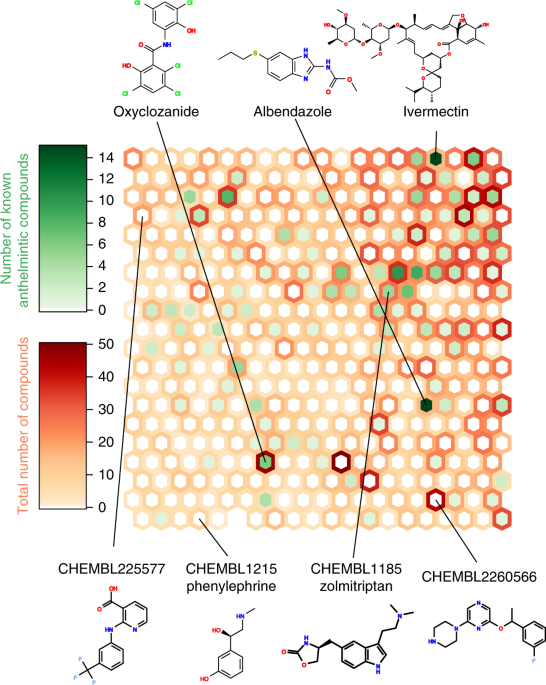
21d). We used a self-organizing map to cluster these compounds based on their molecular fingerprints (Fig.
6).
This classification showed that the screening set was significantly
more structurally diverse than existing anthelmintic compounds
(Supplementary Fig.
24).
The
289 targets were further reduced to 40 high-priority targets, based on
predicted selectivity, avoidance of side-effects (clade-specific
chokepoints or lack of human homologues) and putative vulnerabilities,
such as those suggested by gene family expansions in parasite lineages,
or belonging to pathways containing known or likely anthelmintic targets
(Supplementary Fig.
25).
These 40 targets were associated with 720 drug-like compounds
comprising 181 phase III/IV drugs and 539 medicinal chemistry compounds.
There is independent evidence that some of these have anthelmintic
activity. For example, we identified several compounds that potentially
target glycogen phosphorylase, which is in the same pathway as a likely
anthelmintic target (glycogen phosphorylase phosphatase, likely target
of niridazole; Supplementary Fig.
25). These compounds included the phase III drug alvocidib (flavopiridol), which has anthelmintic activity against
C. elegans96. Another example is the target cathepsin B, expanded in nematode clade Va (Supplementary Table
9a),
for which we identified several compounds including the phase III drug
odanacatib, which has been shown to have anthelmintic activity against
hookworms
97.
Existing drugs such as these are attractive candidates for repurposing
and fast-track therapy development, while the medicinal chemistry
compounds provide a starting point for broader anthelmintic screening.
Discussion
The
evolution of parasitism in nematodes and platyhelminths occurred
independently, starting from different ancestral gene sets and
physiologies. Despite this, common selective pressures of adaptation to
host gut, blood or tissue environments, the need to avoid hosts’ immune
systems, and the acquisition of complex life cycles to effect
transmission, may have driven adaptations in common biological pathways.
While previous comparative analyses of parasitic worms have been
limited to a small number of species within narrow clades, we have
surveyed parasitic worms spanning two phyla, with a focus on those
infecting humans and livestock. A large body of draft genome data (both
published and unpublished) was utilized but, by focusing on
lineage-specific trends rather than individual species-specific
differences, our analysis was deliberately conservative. In particular,
we have focused on large gene family expansions, supported by the
best-quality data and for which functional information was available.
Sequencing of further free-living species, better functional
characterization, and identification of remote orthologs (particularly
for platyhelminths
87),
will undoubtedly refine the resolution of parasite-specific
differences, but our gene family analyses have already revealed
expansions and synapomorphies in functional classes of likely importance
to parasitism, such as feeding and interaction with hosts. We have used
a drug repurposing approach to predict potential new anthelmintic drug
targets and drugs/drug-like compounds that we urge the community to
explore. Further new potential drug targets, worthy of high-throughput
compound screening, may be exposed by the losses of key metabolic
pathways and horizontally acquired genes that we find in particular
parasite groups. This is an unprecedented dataset of parasitic worm
genomes that provides a new type of pan-species reference and a much
needed stimulus to the study of parasitic worm biology.
URLs
SMALT,
http://www.sanger.ac.uk/science/tools/smalt-0; RepeatModeler,
http://www.repeatmasker.org/RepeatModeler.html; TransposonPSI,
http://transposonpsi.sourceforge.net; RepeatMasker,
http://www.repeatmasker.org; code for calculating gene family metrics,
http://tinyurl.com/comparaFamiliesAnalysis-py; WormBase ParaSite,
https://parasite.wormbase.org/.
Methods
Sample collection and preparation
Sources of material and sequencing approaches are summarized in Supplementary Table
1.
Wellcome Sanger Institute (WSI) data production
The genomes of 36 species (Supplementary Tables
1 and
2) were sequenced at WSI. The
C. elegans N2 was also resequenced at WSI.
WSI sequencing and assembly
PCR-free 400–550 bp paired-end Illumina libraries were prepared from <0 .1="" 5="" as="" described="" dna="" for="" g="" genomic="" i="" ng="" to="">Strongyloides stercoralis
18. Where there was insufficient DNA, adapter-ligated material was subjected to ~8 PCR cycles.We used 1–10 μg gDNA or whole genome amplification DNA to generate 3 kb mate-pair libraries, as described for
S. stercoralis18.
If there was insufficient gDNA, whole genome amplification was
performed using GenomiPhi v2. Each library was run on ≥1 Illumina HiSeq
2000 lane.
Short insert paired-end reads were corrected and assembled with SGA v0.9.7
98 (Supplementary Fig.
26a). This assembly was used to calculate the
k-mer distribution for all odd
k of 41–81, using GenomeTools v.1.3.7
99. The
k-mer length for which the maximum number of unique
k-mers was present was used as the
k-mer setting in a second assembly, using Velvet v1.2.03
100 with SGA-corrected reads. For species with 3 kb mate-pair data, the Velvet assembly was scaffolded using SSPACE
101. Contigs were extended, and gaps closed and shortened, using Gapfiller
102 and IMAGE
103. Short fragment reads were remapped to the assembly using SMALT (see URLs), and unaligned reads assembled using Velvet
100 and this merged with the main assembly. The assembly was re-scaffolded using SSPACE
101, and consensus base quality improved with iCORN
104. REAPR
105 was used to break incorrectly assembled scaffolds/contigs. We carried out manual improvement for
Wuchereria bancrofti and
D. medinensis using Gap5
106 and Illumina read-pairs.
WSI assembly quality control
Contamination screening. Assemblies were screened for contamination using BLAST
107 against vertebrate and invertebrate sequences (see ref.
108). For
Anisakis simplex, the assembly contained minor laboratory contamination with
S. mansoni, which we removed using BLASTN against
S. mansoni.
Assembly completeness. CEGMA v2.4
109
was used to assess completeness. Consistent sets of CEGMA genes were
missing from some phylogenetic groups (Supplementary Table
2); these were discounted from the completeness calculation for those species (‘CEGMA’ in Supplementary Table
2).
Effect
of repeats. We re-mapped the short-insert library’s reads to the
appropriate assembly using SMALT (see URLs; indexing -k13 -s4 and
mapping -y 0.9 -x -r 1). For each scaffold of ≥8 kb, median (med
s) and mean (
ms) per-base read-depth were calculated using BEDTools
110, and genome-wide depth (med
g) calculated as the median med
s (ref.
17). For a
ls bp scaffold, the extra sequence that would be gained by ‘uncollapsing’ repeats was estimated as
es = (
ms − med
g) ×
ls/med
g (Supplementary Table
5).
WSI gene prediction
Our pipeline
111 had four steps (Supplementary Fig.
27a). First, repeats were masked. Second, preliminary gene predictions, to use as input for MAKER v2.2.28
112 were generated using Augustus 2.5.5
113, SNAP 2013-02-16
114, GeneMark-ES 2.3a
115, genBlastG
116 and RATT
117. Third, species-specific ESTs and complementary DNAs from INSDC
118, and proteins from related species, were aligned to the genome using BLAST
107. Last, EST/protein alignments and gene models were used by MAKER to produce a gene set.
McDonnell Genome Institute (MGI) data production
The genomes of six species were sequenced at MGI (Supplementary Tables
1 and
2).
MGI sequencing, assembly and quality control
Genome sequencing was carried out on Illumina and 454 instruments (see ref.
119). The workflow for each assembly is in Supplementary Table
1.
Three
kilobase, 8 kb and fragment 454 reads (or Illumina reads) were subject
to adapter removal, quality trimming and length filtering (Supplementary
Fig.
26b). Cleaned 454 reads were assembled using Newbler
120
before being scaffolded with an in-house tool CIGA, which links contigs
based on cDNA evidence. Cleaned Illumina reads were assembled using
AllPaths-LG
121.
The assembly was scaffolded further using an in-house tool Pygap, using
Illumina short paired-end sequences; and L_RNA_scaffolder
122, using 454 cDNA data.
An assisted assembly approach was used for
Trichinella nativa, whereby ‘cleaned’ Illumina 3 kb paired-end sequence data were mapped against the
T. spiralis genome using bwa
123 (Supplementary Fig.
26b), and the
T. nativa residues were substituted at aligned positions (see ref.
119).
Adaptor sequences and contaminants were identified by comparison to a database of vectors and contaminants, using Megablast
124.
MGI transcriptome sequencing and gene prediction
Transcriptome libraries (Supplementary Table
22) were generated with the Illumina TS stranded protocol, and reads assembled using Trinity
125 (see ref.
119).
Genes were predicted using MAKER
112, based on input gene models from SNAP
114, FGENESH (Softberry), Augustus
113, and aligned messenger RNA, EST, transcriptome and protein data from the same or related species (Supplementary Fig.
27b; see ref.
119).
Blaxter Nematode and Neglected Genomics (BaNG) data production
The genomes of three species were sequenced by BaNG (Supplementary Tables
1 and
2).
Sequencing
was performed on Illumina HiSeq 2000 and HiSeq 2500 instruments, using
100 or 125 base, paired-end protocols. Paired-end libraries were
generated using the Illumina TruSeq protocol.
Sequence data were filtered of contaminating host reads using blobtools
126. Cleaned reads were normalized with the khmer software
127 using a
k-mer of 41, and then assembled with ABySS (v1.3.3)
128, with a minimum of three pairs needed to connect contigs during scaffolding (
n = 3) (Supplementary Fig.
26c). Assemblies were assessed using blobtools and CEGMA
109.
Augustus
113 was used to predict gene models, trained using annotations from MAKER
112. As hints for MAKER, we used
Litomosoides sigmodontis 454 RNA sequencing data assembled with MIRA
129 and Newbler
120, and
Onchocerca ochengi Illumina RNA sequencing data
130 assembled using Trinity
131 (Supplementary Fig.
27c).
Defining high-quality ‘tier 1’ species
A
subset of nematode and platyhelminth genomes, termed ‘tier 1’, was
selected that had better-quality assemblies and spanned the major clades
(Supplementary Table
4).
To choose these, species were selected that (1) had contiguous
assemblies (usually N50/scaffold-count >5), and complete proteomes
(usually CEGMA partial >85%), or (2) that helped to ensure ~50% of
the genera in each species group (‘Analysis group’ in Supplementary
Table
4) were represented.
Analysis of repeat content and genome size
For each species, repeat libraries were built using RepeatModeler (see URLs), TransposonPSI (see URLs) and LTRharvest
132, and the three libraries merged (see ref.
133). The merged library was used to mask repeats in a species’ genome using RepeatMasker (see URLs; –s).
The
initial standard regression model and stepwise model fitting used ‘lm’
and ‘step’ in R v3.2.2. The Bayesian mixed-effect model used MCMCglmm
134 (v2.24). To create a mixed-effect model, the species tree (see
Methods) was transformed into an ultrametric tree using PATHd8
135,
with a small constant added to short branches to ensure no zero-length
branches were reconstructed; and outgroup species were removed.
Compara database
An in-house Ensembl Compara
46 database was constructed containing the 81 platyhelminths and nematodes, and 10 additional outgroups (Supplementary Table
2). All parasitic nematode/platyhelminth species with gene sets available at the time (April 2014) were included.
The
species tree used to construct the initial version of our database used
an edited version of the National Center for Biotechnology Information
(NCBI) taxonomy
136
with several controversial speciation nodes represented as
multifurcations. For our final database, the input species tree was
derived by building a tree based on the previous database version, based
on one-to-one orthologs present in ≥20 species. To do this, proteins in
each ortholog group were aligned using MAFFT v6.857
137; alignments trimmed using GBlocks v0.91b
138, concatenated and used to build a maximum likelihood tree using a partitioned analysis in RAxML v7.8.6
139, using the minimum Akaike’s information criterion (minAIC) model for each ortholog group.
The database was queried to identify gene families, orthologs and paralogs.
Species tree and tree based on gene family presence
We identified 202 gene families present in ≥25% of the 91 species (81 helminths and 10 outgroups) in our Compara database (
Methods) and always single-copy. For each family, amino acid sequences were aligned using MAFFT v7.205
137 (-auto). Each alignment was trimmed using GBlocks v0.91b
138
(-b4 = 4 -b3 = 4 -b5 = h), and its likelihood calculated on a
maximum-parsimony guide tree for all relatively simple (single-matrix)
amino acid substitution models in RAxML v8.0.24
139,
and the minAIC model identified. Alignments were concatenated and a
maximum-likelihood tree built, under a partitioned model in which sites
from a gene were assigned the minAIC model for that gene, with a
discrete gamma distribution of rates across sites. Relationships within
outgroup lineages were constrained to match the standard view of
metazoan relationships (for example, Dunn et al.
140).
The final tree was the highest likelihood one from five search
replicates with different random number seeds. One hundred bootstrap
resampling replicates were performed, each based on a single rapid
search.
We also constructed a maximum-likelihood phylogeny based
on gene family presence/absence for families not shared by all 81
nematode/platyhelminth species, using RAxML v8.2.8
139, with a two-state model and the Lewis method to correct for absence of constant-state observations.
Functional annotation
InterProScan
141
v5.0.7 was used to identify conserved domains from all predicted
proteins. A name was assigned to each predicted protein based on curated
information in UniProt
142 for orthologs identified from our Compara database (
Methods), or based on InterPro
143 domains (see ref.
144). Gene ontology (GO) terms were assigned by transferring GO terms from orthologs
144, and using InterProScan.
Signal peptides and transmembrane domains were predicted using Phobius
145 v1.01 and SecretomeP
146
v1.0. A protein predicted by Phobius to have a transmembrane domain was
categorized as ‘membrane-bound’, and non-membrane-bound proteins as
‘classically secreted’ if Phobius predicted a signal peptide within 70
amino acids of their start. Remaining proteins in which SecretomeP
predicted a signal peptide were classified as ‘non-classically secreted’
(Supplementary Table
7).
Pairwise
combinations of Pfam domains were identified in proteins of the 81
nematodes and platyhelminths. After excluding those present in complete
genomes of other phyla in UniProt (June 2016), we classified a
combination as ‘nematode-specific’ (or ‘flatworm-specific’) if it was
present in >30% of nematodes (platyhelminths) and no platyhelminths
(nematodes) (Supplementary Table
14).
Synapomorphic gene families
Families in our Compara database (
Methods) were analyzed using KinFin v0.8.3
147, by providing InterPro IDs (
Methods) and a species tree that had clades III, IV and V as a polytomy (Fig.
2). Synapomorphic families were identified at 25 nodes of interest (Supplementary Table
8),
by using Dollo parsimony and requiring a family must contain genes from
≥1 descendant species from each child node of the node of interest, and
must not contain other species. Families were filtered to retain those
that (1) contained ≥90% of descendant species of the node of interest,
and (2) in which >90% of species contained ≥1 gene with a particular
InterPro domain.
Candidate lateral gene transfers
Ferrochelatase families in our Compara database (
Methods)
were extracted by screening for a Ferrochelatase (IPR001015) domain.
Additional ferrochelatases were retrieved from NCBI for 17 bacterial
taxa (Supplementary Table
8c). Sequences were aligned using MAFFT v7.267 (E-INS-i algorithm)
137 and the alignment trimmed using trimAl v1.4
148. Phylogenetic analysis was carried out using RAxML
139
under the PROTGAMMAGTR model, and 20 alternative runs on distinct
starting trees. Non-parametric bootstrap analysis was carried out for
100 replicates.
For cobyric acid synthase and acetate/succinate
transporter, the top BLAST hits from GenBank, and representative
sequences from other taxonomic groups, were aligned with MAFFT v7.205
137 (-auto), and alignments trimmed with trimal v1.4
148. Phylogenetic analyses were performed using RAxML v8.2.8
139
under the model that minimized the AIC (LG4X for cobyric acid synthase,
LG4M for acetate transporter), based on 5 random-addition-sequence
replicates, and 100 non-parametric bootstrap replicates.
Gene family expansions
We used three metrics to identify families in our Compara database (
Methods) that varied greatly in gene count across species (see ref.
149).
To control for fragmented assemblies, we used summed protein length per
species (in a family) as a proxy for gene count in these metrics:
1. Coefficient of variation:
𝑐.𝑣.=𝑠/𝑥¯
where
s is the standard deviation in summed protein length per species, and
𝑥¯
its mean.
2. Maximum
Z-score:
𝑍max=max𝑐∈𝑇(𝑥¯𝑖,𝑖∈𝑐−𝑥¯𝑠𝑖,𝑖∉𝑐)
where
T is the set of non-overlapping species groups (‘Analysis group’ in Supplementary Table
4),
c a group in
T, index
i refers to a particular species,
𝑥¯𝑖,𝑖∈𝑐
the mean of the summed protein length (per species) in
c, and
𝑠𝑖,𝑖∉𝑐 the standard deviation in summed protein length per species in species outside
c.
3. Maximum enrichment coefficient:
𝐸max=max𝑐∈𝑇(𝑥¯𝑖,𝑖∈𝑐𝑥¯𝑖,𝑖∉𝑐)
To increase reliability, these metrics were calculated by only considering tier 1 species (those with high-quality assemblies;
Methods). Our code for calculating metrics is available (see URLs).
SCP/TAPS
SCP/TAPS genes were identified as having Pfam PF00188, or being in a SCP/TAPS family in our Compara database (
Methods). Those between 146 aa (shortest
C. elegans SCP/TAPS) and 1,000 aa were included in the phylogenetic analysis (Supplementary Table
10). Clusters were detected among sequences from a species group (‘analysis group’ in Supplementary Table
4) using USEARCH
150
(UCLUST, aa identity cut-off = 0.70), and a consensus sequence
generated for each cluster. The consensus sequences were aligned using
MAFFT
137 (v7.271, –localpair –maxiterate 2 –retree 1 –bl 45); the alignment trimmed with trimAl
148 (-gt 0.006); and a maximum likelihood tree built using FastTreeMP
151 (v2.1.7 SSE3, -wag -gamma).
Proteins historically targeted for drug development
Each nematode/platyhelminth proteome was searched against candidate proteases using MEROPS batch-BLAST
152 (
E < 0.001), and PfamScan
153 was used to identify additional homologues in some species (Supplementary Table
11).
Putative GPCRs, identified from the literature and GO:0004930 annotations in WormBase
154, were used to identify families in our Compara database (
Methods). For each family, HHSuite
155
was used to search Uniprot, SCOPUS, Pfam, and PDB; 200 families hitting
≥2 databases were deemed actual GPCR families (see ref.
156). Additional families were identified from synapomorphies (
Methods) and curation, giving 230 GPCR families (Supplementary Table
15).
To build a phylogenetic tree of ion channels, known genes from
C. elegans157,
Brugia malayi158,
Haemonchus contortus159,
Oesophagostomum dentatum159 and
S. mansoni84 were gathered, and their homologues in Compara families in WormBase ParaSite
160. Genes with <3 or="">8 transmembrane domains (predicted by HMMTOP
161) were discarded. Genes were aligned with MAFFT
137, and the alignment trimmed with trimAl
148. The phylogeny was inferred with MrBayes3.2
162.
Posterior probabilities were calculated from eight reversible jump
Markov chain Monte Carlo chains over 20,000,000 generations.
Kinase models were taken from Kinomer
163, and thresholds optimized to detect known
C. elegans kinases (see ref.
164).
The final thresholds were used to filter HMMER search results (against
Kinomer) for nematode and platyhelminth species (Supplementary Table
23).
C. elegans ABC transporter and cys-loop receptor subunit genes were collated from WormBase
154, to which we added
H. contortus acr-26 and
acr-27 (absent from
C. elegans85). Homologs in nematodes and platyhelminths were identified using BLASTP (Supplementary Tables
16 and
17).
GO and InterPro/Pfam annotation enrichment
Counts
of proteins annotated with each GO term (or InterPro/Pfam domain) per
species were normalized by dividing by the total GO annotations in a
particular species. To test for enrichment of a particular GO term in a
species group (‘analysis group’ in Supplementary Table
4), we used a Mann-Whitney
U test to compare normalized counts in that species group, to those in all other species (Supplementary Table
24).
Metabolism
EC (Enzyme Commission number) predictions for nematodes and platyhelminths were derived by combining DETECT v2.0
165, PRIAM
166, KAAS
167 and BRENDA
168 (see ref.
169, Supplementary Fig.
28 and Supplementary Table
18), and supplemented for the 33 tier 1 species (
Methods) by pathway hole-filling using Pathway Tools
170 (v18.5). Comparisons of all 81 species (Supplementary Fig.
20a and Supplementary Table
20) did not include ECs from hole-filling. Lower confidence ECs were inferred using families from our Compara database (
Methods). Auxotrophies were predicted using Pathway Tools and BioCyc
171. To predict carbohydrate-active enzymes, HMMER3 was used to search dbCAN
172 (Supplementary Table
25).
Pathway coverage was the fraction of ECs in a reference pathway that were annotated in a species (see ref.
173). We included pathways for which KEGG had a reference pathway for a nematode/platyhelminth (Supplementary Table
18e). Presence of KEGG modules was predicted using modDFS
174, and species clustered based on module presence using Ward-linkage, based on Jaccard similarity index
175.
Chokepoint enzymes were predicted following Taylor et al.
176, using subnetworks of KEGG networks formed by just the enzymes (ECs) we had annotated in each particular species.
Potential anthelmintic drug targets and drugs
Potential drug targets
Nematode and platyhelminth proteins from tier 1 species (with high-quality assemblies;
Methods) were searched against single-protein targets from ChEMBL v21
177 using BLASTP (
E ≤ 1 × 10
−10). After collapsing by gene family, 1,925 worm genes remained.
To
assign a ‘target score’ to each worm gene, the main factors considered
were similarity to known drug targets; lack of human homologues; and
whether
C. elegans/
Drosophila melanogaster homologues had lethal phenotypes (see ref.
178).
Potential new anthelmintic drugs
ChEMBL v21
177
was used to identify 827,889 compounds with activities against ChEMBL
targets to which worm proteins had BLAST matches. To calculate ‘compound
scores’, we prioritized compounds in high clinical development phases,
oral/topical administration, crystal structures, properties consistent
with oral drugs and lacking toxicity (see ref.
178).
Our
top 15% (249) of highest-scoring worm targets had 292,499 compounds.
These were filtered by selecting compounds that (1) co-appeared in a
PDBe
179
(Protein Data Bank in Europe) structure with the ChEMBL target; or (2)
had median pChEMBL > 5; leaving 131,452 ‘top drug candidates’.
A ‘diverse screening set’
The 131,452 candidates were placed into 27,944 chemical classes, based on ECFP4 fingerprints (see ref.
178).
They were filtered by (1) discarding medicinal chemistry compounds that
did not co-appear in a PDBe structure with the ChEMBL target, or have
median pCHEMBL > 7; (2) checking availability for purchase in ZINC 15
180; and (3) for each worm target, taking the highest-scoring compound from each class; this gave 5,046 compounds.
Self-organizing map
We constructed a self-organizing map of our diverse screening set plus known anthelmintic compounds (Supplementary Table
21a; see ref.
178), using Kohonen v3.02
181
in R v3.3.0, using a 20 × 20 cell hexagonal, non-toroidal grid. The
self-organizing map was trained for 4,000 steps, where training
optimized Tanimoto distances between ECFP4 fingerprints.
Reporting Summary
Further information on research design is available in the
Nature Research Reporting Summary linked to this article.
Data availability
Sequence data have been deposited in the European
Nucleotide Archive (ENA). Assemblies and annotation are available at
WormBase and WormBase-ParaSite (
https://parasite.wormbase.org/). All have been submitted to GenBank under the BioProject IDs listed in Supplementary Table
1.
Publisher’s note: Springer Nature remains neutral with regard to jurisdictional claims in published maps and institutional affiliations.
References
- 1.
G.B.D.
2015 Disease and Injury Incidence and Prevalence Collaborators. Global,
regional, and national incidence, prevalence, and years lived with
disability for 310 diseases and injuries, 1990-2015: a systematic
analysis for the Global Burden of Disease Study 2015. Lancet 388, 1545–1602 (2016).
2.
Charlier,
J., van der Voort, M., Kenyon, F., Skuce, P. & Vercruysse, J.
Chasing helminths and their economic impact on farmed ruminants. Trends Parasitol. 30, 361–367 (2014).
3.
Jones, J. T. et al. Top 10 plant-parasitic nematodes in molecular plant pathology. Mol. Plant Pathol. 14, 946–961 (2013).
4.
Furtado, L. F., de Paiva Bello, A. C. & Rabelo, E. M. Benzimidazole resistance in helminths: From problem to diagnosis. Acta Trop. 162, 95–102 (2016).
5.
Kaplan, R. M. & Vidyashankar, A. N. An inconvenient truth: global worming and anthelmintic resistance. Vet. Parasitol. 186, 70–78 (2012).
6.
Hewitson, J. P. & Maizels, R. M. Vaccination against helminth parasite infections. Expert Rev. Vaccines 13, 473–487 (2014).
7.
Ntalli, N. G. & Caboni, P. Botanical nematicides: a review. J. Agric. Food. Chem. 60, 9929–9940 (2012).
8.
Young, N. D. et al. Whole-genome sequence of Schistosoma haematobium. Nat. Genet. 44, 221–225 (2012).
9.
The Schistosoma japonicum Genome Sequencing and Functional Analysis Consortium. The Schistosoma japonicum genome reveals features of host–parasite interplay. Nature 460, 345–351 (2009).
10.
Protasio,
A. V. et al. A systematically improved high quality genome and
transcriptome of the human blood fluke Schistosoma mansoni. PLoS Negl. Trop. Dis. 6, e1455 (2012).
11.
Wang, X. et al. The draft genome of the carcinogenic human liver fluke Clonorchis sinensis. Genome. Biol. 12, R107 (2011).
12.
McNulty, S. N. et al. Genomes of Fasciola hepatica from the Americas reveal colonization with Neorickettsia endobacteria related to the agents of potomac horse and human sennetsu fevers. PLoS Genet. 13, e1006537 (2017).
13.
Tsai, I. J. et al. The genomes of four tapeworm species reveal adaptations to parasitism. Nature 496, 57–63 (2013).
14.
Bennett, H. M. et al. The genome of the sparganosis tapeworm Spirometra erinaceieuropaei isolated from the biopsy of a migrating brain lesion. Genome. Biol. 15, 510 (2014).
15.
Schiffer, P. H. et al. The genome of Romanomermis culicivorax: revealing fundamental changes in the core developmental genetic toolkit in Nematoda. BMC Genomics 14, 923 (2013).
16.
Mitreva, M. et al. The draft genome of the parasitic nematode Trichinella spiralis. Nat. Genet. 43, 228–235 (2011).
17.
Foth,
B. J. et al. Whipworm genome and dual-species transcriptome analyses
provide molecular insights into an intimate host–parasite interaction. Nat. Genet. 46, 693–700 (2014).
18.
Hunt, V. L. et al. The genomic basis of parasitism in the Strongyloides clade of nematodes. Nat. Genet. 48, 299–307 (2016).
19.
Kikuchi, T. et al. Genomic insights into the origin of parasitism in the emerging plant pathogen Bursaphelenchus xylophilus. PLoS Pathog. 7, e1002219 (2011).
20.
Cotton, J. A. et al. The genome and life-stage specific transcriptomes of Globodera pallida elucidate key aspects of plant parasitism by a cyst nematode. Genome Biol. 15, R43 (2014).
21.
Opperman, C. H. et al. Sequence and genetic map of Meloidogyne hapla: a compact nematode genome for plant parasitism. Proc. Natl Acad. Sci. USA 105, 14802–14807 (2008).
22.
Ghedin, E. et al. Draft genome of the filarial nematode parasite Brugia malayi. Science 317, 1756–1760 (2007).
23.
Godel, C. et al. The genome of the heartworm, Dirofilaria immitis, reveals drug and vaccine targets. FASEB J. 26, 4650–4661 (2012).
24.
Desjardins, C. A. et al. Genomics of Loa loa, a Wolbachia-free filarial parasite of humans. Nat. Genet. 45, 495–500 (2013).
25.
Cotton, J. A. et al. The genome of Onchocerca volvulus, agent of river blindness. Nat. Microbiol. 2, 16216 (2016).
26.
Wang, J. et al. Silencing of germline-expressed genes by DNA elimination in somatic cells. Dev. Cell 23, 1072–1080 (2012).
27.
Tang, Y. T. et al. Genome of the human hookworm Necator americanus. Nat. Genet. 46, 261–269 (2014).
28.
Tyagi,
R. et al. Cracking the nodule worm code advances knowledge of parasite
biology and biotechnology to tackle major diseases of livestock. Biotechnol. Adv. 33, 980–991 (2015).
29.
McNulty, S. N. et al. Dictyocaulus viviparus genome, variome and transcriptome elucidate lungworm biology and support future intervention. Sci. Rep. 6, 20316 (2016).
30.
Laing, R. et al. The genome and transcriptome of Haemonchus contortus, a key model parasite for drug and vaccine discovery. Genome. Biol. 14, R88 (2013).
31.
C. elegans Sequencing Consortium. Genome sequence of the nematode C. elegans: a platform for investigating biology. Science 282, 2012–2018 (1998).
32.
Dieterich, C. et al. The Pristionchus pacificus genome provides a unique perspective on nematode lifestyle and parasitism. Nat. Genet. 40, 1193–1198 (2008).
33.
Robb, S. M., Ross, E. & Sanchez Alvarado, A. SmedGD: the Schmidtea mediterranea genome database. Nucleic Acids Res. 36, D599–D606 (2008).
34.
Srinivasan, J. et al. The draft genome and transcriptome of Panagrellus redivivus are shaped by the harsh demands of a free-living lifestyle. Genetics 193, 1279–1295 (2013).
35.
Srivastava, M. et al. The Amphimedon queenslandica genome and the evolution of animal complexity. Nature 466, 720–726 (2010).
36.
Simakov, O. et al. Insights into bilaterian evolution from three spiralian genomes. Nature 493, 526–531 (2013).
37.
Satou, Y. et al. Improved genome assembly and evidence-based global gene model set for the chordate Ciona intestinalis: new insight into intron and operon populations. Genome Biol. 9, R152 (2008).
38.
Zhang, G. et al. The oyster genome reveals stress adaptation and complexity of shell formation. Nature 490, 49–54 (2012).
39.
Howe, K. et al. The zebrafish reference genome sequence and its relationship to the human genome. Nature 496, 498–503 (2013).
40.
Adams, M. D. et al. The genome sequence of Drosophila melanogaster. Science 287, 2185–2195 (2000).
41.
International Human Genome Sequencing Consortium. Initial sequencing and analysis of the human genome. Nature 409, 860–921 (2001).
42.
Pagel Van Zee, J. et al. Tick genomics: the Ixodes genome project and beyond. Int. J. Parasitol. 37, 1297–1305 (2007).
43.
Putnam, N. H. et al. Sea anemone genome reveals ancestral eumetazoan gene repertoire and genomic organization. Science 317, 86–94 (2007).
44.
Srivastava, M. et al. The Trichoplax genome and the nature of placozoans. Nature 454, 955–960 (2008).
45.
Lynch, M., Bobay, L. M., Catania, F., Gout, J. F. & Rho, M. The repatterning of eukaryotic genomes by random genetic drift. Annu. Rev. Genomics Hum. Genet. 12, 347–366 (2011).
46.
Vilella, A. J. et al. EnsemblCompara GeneTrees: complete, duplication-aware phylogenetic trees in vertebrates. Genome Res. 19, 327–335 (2009).
47.
Rao, A. U., Carta, L. K., Lesuisse, E. & Hamza, I. Lack of heme synthesis in a free-living eukaryote. Proc. Natl Acad. Sci. USA 102, 4270–4275 (2005).
48.
Wu, B. et al. Interdomain lateral gene transfer of an essential ferrochelatase gene in human parasitic nematodes. Proc. Natl Acad. Sci. USA 110, 7748–7753 (2013).
49.
Nagayasu, E. et al. Identification of a bacteria-like ferrochelatase in Strongyloides venezuelensis, an animal parasitic nematode. PLoS ONE 8, e58458 (2013).
50.
Casaravilla, C. et al. Characterization of myo-inositol hexakisphosphate deposits from larval Echinococcus granulosus. FEBS J. 273, 3192–3203 (2006).
51.
Diaz, A., Casaravilla, C., Barrios, A. A. & Ferreira, A. M. Parasite molecules and host responses in cystic echinococcosis. Parasite Immunol. 38, 193–205 (2016).
52.
Santos, R. et al. A comprehensive map of molecular drug targets. Nat. Rev. Drug Discov. 16, 19–34 (2017).
53.
Valentim,
C. L. et al. Genetic and molecular basis of drug resistance and
species-specific drug action in schistosome parasites. Science 342, 1385–1389 (2013).
54.
Parsons, L. M. et al. Caenorhabditis elegans bacterial pathogen resistant bus-4 mutants produce altered mucins. PLoS ONE 9, e107250 (2014).
55.
Shapira, M. et al. A conserved role for a GATA transcription factor in regulating epithelial innate immune responses. Proc. Natl Acad. Sci. USA 103, 14086–14091 (2006).
56.
Hewitson, J. P. et al. Proteomic analysis of secretory products from the model gastrointestinal nematode Heligmosomoides polygyrus reveals dominance of venom allergen-like (VAL) proteins. J. Proteomics 74, 1573–1594 (2011).
57.
van
der Hoeven, R., Cruz, M. R., Chavez, V. & Garsin, D. A.
Localization of the dual oxidase BLI-3 and characterization of its NADPH
oxidase domain during infection of Caenorhabditis elegans. PLoS ONE 10, e0124091 (2015).
58.
Esteban,
M. R., Giovinazzo, G., de la Hera, A. & Goday, C. PUMA1: a novel
protein that associates with the centrosomes, spindle and centromeres in
the nematode Parascaris. J. Cell Sci. 111, 723–735 (1998).
59.
Tobler, H., Etter, A. & Muller, F. Chromatin diminution in nematode development. Trends Genet. 8, 427–432 (1992).
60.
Albarqi, M. M. et al. Regulation of life cycle checkpoints and developmental activation of infective larvae in Strongyloides stercoralis by dafachronic acid. PLoS Pathog. 12, e1005358 (2016).
61.
Zarlenga, D. S., Nisbet, A. J., Gasbarre, L. C. & Garrett, W. M. A calcium-activated nucleotidase secreted from Ostertagia ostertagi 4th-stage larvae is a member of the novel salivary apyrases present in blood-feeding arthropods. Parasitology 138, 333–343 (2011).
62.
Cathcart,
M. K. & Bhattacharjee, A. Monoamine oxidase A (MAO-A): a signature
marker of alternatively activated monocytes/macrophages. Inflamm. Cell Signal. 1, e161 (2014).
63.
Coakley,
G., Maizels, R. M. & Buck, A. H. Exosomes and other extracellular
vesicles: the new communicators in parasite infections. Trends Parasitol. 31, 477–489 (2015).
64.
Wu, C. et al. Mapping the binding between the tetraspanin molecule (Sjc23) of Schistosoma japonicum and human non-immune IgG. PLoS ONE 6, e19112 (2011).
65.
Krautz-Peterson, G. et al. Schistosoma mansoni
infection of mice, rats and humans elicits a strong antibody response
to a limited number of reduction-sensitive epitopes on five major
tegumental membrane proteins. PLoS Negl. Trop. Dis. 11, e0005306 (2017).
66.
Prior, A. et al. A surface-associated retinol- and fatty acid-binding protein (Gp-FAR-1) from the potato cyst nematode Globodera pallida: lipid binding activities, structural analysis and expression pattern. Biochem. J. 356, 387–394 (2001).
67.
Rey-Burusco,
M. F. et al. Diversity in the structures and ligand-binding sites of
nematode fatty acid and retinol-binding proteins revealed by Na-FAR-1
from Necator americanus. Biochem. J. 471, 403–414 (2015).
68.
Dell, A., Haslam, S. M. & Morris, H. R. in Parasitic Nematodes: Molecular Biology, Biochemistry and Immunology (eds. Kennedy, M. W. & Harnett, W.) 285–307 (Cabi Publishing, Oxfordshire, UK, 2013).
69.
Rodrigues, J. A. et al. Parasite glycobiology: a bittersweet symphony. PLoS Pathog. 11, e1005169 (2015).
70.
Anderson, L. et al. Schistosoma mansoni egg, adult male and female comparative gene expression analysis and identification of novel genes by RNA-Seq. PLoS Negl. Trop. Dis. 9, e0004334 (2015).
71.
Gong, H. et al. A novel PAN/apple domain-containing protein from Toxoplasma gondii: characterization and receptor identification. PLoS ONE 7, e30169 (2012).
72.
Cantacessi,
C. et al. A portrait of the “SCP/TAPS” proteins of
eukaryotes—developing a framework for fundamental research and
biotechnological outcomes. Biotechnol. Adv. 27, 376–388 (2009).
73.
McKerrow, J. H., Caffrey, C., Kelly, B., Loke, P. & Sajid, M. Proteases in parasitic diseases. Annu. Rev. Pathol. 1, 497–536 (2006).
74.
Williamson, A. L. et al. Ancylostoma caninum MTP-1, an astacin-like metalloprotease secreted by infective hookworm larvae, is involved in tissue migration. Infect. Immun. 74, 961–967 (2006).
75.
Williamson, A. L. et al. A multi-enzyme cascade of hemoglobin proteolysis in the intestine of blood-feeding hookworms. J. Biol. Chem. 279, 35950–35957 (2004).
76.
Delcroix, M. et al. A multienzyme network functions in intestinal protein digestion by a platyhelminth parasite. J. Biol. Chem. 281, 39316–39329 (2006).
77.
Duffy,
M. S., Cevasco, D. K., Zarlenga, D. S., Sukhumavasi, W. & Appleton,
J. A. Cathepsin B homologue at the interface between a parasitic
nematode and its intermediate host. Infect. Immun. 74, 1297–1304 (2006).
78.
Cancela, M. et al. A distinctive repertoire of cathepsins is expressed by juvenile invasive Fasciola hepatica. Biochimie 90, 1461–1475 (2008).
79.
Knox, D. P. Proteinase inhibitors and helminth parasite infection. Parasite Immunol. 29, 57–71 (2007).
80.
Rehman, A. A., Ahsan, H. & Khan, F. H. Alpha-2-macroglobulin: a physiological guardian. J. Cell. Physiol. 228, 1665–1675 (2013).
81.
Martzen, M. R., Geise, G. L., Hogan, B. J. & Peanasky, R. J. Ascaris suum: localization by immunochemical and fluorescent probes of host proteases and parasite proteinase inhibitors in cross-sections. Exp. Parasitol. 60, 139–149 (1985).
82.
Nei,
M., Niimura, Y. & Nozawa, M. The evolution of animal chemosensory
receptor gene repertoires: roles of chance and necessity. Nat. Rev. Genet. 9, 951–963 (2008).
83.
Lynagh, T. et al. Molecular basis for convergent evolution of glutamate recognition by pentameric ligand-gated ion channels. Sci. Rep. 5, 8558 (2015).
84.
MacDonald, K. et al. Functional characterization of a novel family of acetylcholine-gated chloride channels in Schistosoma mansoni. PLoS Pathog. 10, e1004181 (2014).
85.
Courtot, E. et al. Functional characterization of a novel class of morantel-sensitive acetylcholine receptors in nematodes. PLoS Pathog. 11, e1005267 (2015).
86.
Vasiliou, V., Vasiliou, K. & Nebert, D. W. Human ATP-binding cassette (ABC) transporter family. Hum. Genomics 3, 281–290 (2009).
87.
Martin-Duran,
J. M., Ryan, J. F., Vellutini, B. C., Pang, K. & Hejnol, A.
Increased taxon sampling reveals thousands of hidden orthologs in
flatworms. Genome Res. 27, 1263–1272 (2017).
88.
Kanehisa,
M., Goto, S., Sato, Y., Furumichi, M. & Tanabe, M. KEGG for
integration and interpretation of large-scale molecular data sets. Nucleic Acids Res. 40, D109–D114 (2012).
89.
Kondrashov,
F. A., Koonin, E. V., Morgunov, I. G., Finogenova, T. V. &
Kondrashova, M. N. Evolution of glyoxylate cycle enzymes in Metazoa:
evidence of multiple horizontal transfer events and pseudogene
formation. Biol. Direct. 1, 31 (2006).
90.
Harder, A. The biochemistry of Haemonchus contortus and other parasitic nematodes. Adv. Parasitol. 93, 69–94 (2016).
91.
Marr, J. J. & Müller, M. Biochemistry and Molecular Biology of Parasites (Academic Press, San Diego, CA, USA, 1995).
92.
Pearce, E. J. & Huang, S. C. The metabolic control of schistosome egg production. Cell Microbiol. 17, 796–801 (2015).
93.
Mehlhorn, H. (ed.) Encyclopedia of Parasitology (Springer, New York, NY, USA, 2008).
94.
Watson, E. et al. Metabolic network rewiring of propionate flux compensates vitamin B12 deficiency in C. elegans. eLife 5, e17670 (2016).
95.
Van Soest, P. J. Nutritional Ecology of the Ruminant (Cornell Univ. Press, Ithaca, NY, USA, 1994).
96.
Taylor, C. M. et al. Using existing drugs as leads for broad spectrum anthelmintics targeting protein kinases. PLoS Pathog. 9, e1003149 (2013).
97.
Vermeire,
J. J., Suzuki, B. M. & Caffrey, C. R. Odanacatib, a cathepsin K
cysteine protease inhibitor, kills hookworm in vivo. Pharmaceuticals 9, 39 (2016).
98.
Simpson, J. T. & Durbin, R. Efficient de novo assembly of large genomes using compressed data structures. Genome Res. 22, 549–556 (2012).
99.
Gremme,
G., Steinbiss, S. & Kurtz, S. GenomeTools: a comprehensive software
library for efficient processing of structured genome annotations. IEEE/ACM Trans. Comput. Biol. Bioinform. 10, 645–656 (2013).
100.
Zerbino, D. R. & Birney, E. Velvet: algorithms for de novo short read assembly using de Bruijn graphs. Genome Res. 18, 821–829 (2008).
101.
Boetzer, M., Henkel, C. V., Jansen, H. J., Butler, D. & Pirovano, W. Scaffolding pre-assembled contigs using SSPACE. Bioinformatics 27, 578–579 (2011).
102.
Boetzer, M. & Pirovano, W. Toward almost closed genomes with GapFiller. Genome Biol. 13, R56 (2012).
103.
Tsai,
I. J., Otto, T. D. & Berriman, M. Improving draft assemblies by
iterative mapping and assembly of short reads to eliminate gaps. Genome Biol. 11, R41 (2010).
104.
Otto,
T. D., Sanders, M., Berriman, M. & Newbold, C. Iterative correction
of reference nucleotides (iCORN) using second generation sequencing
technology. Bioinformatics 26, 1704–1707 (2010).
105.
Hunt, M. et al. REAPR: a universal tool for genome assembly evaluation. Genome Biol. 14, R47 (2013).
106.
Bonfield, J. K. & Whitwham, A. Gap5—editing the billion fragment sequence assembly. Bioinformatics 26, 1699–1703 (2010).
107.
Altschul, S. F. et al. Gapped BLAST and PSI-BLAST: a new generation of protein database search programs. Nucleic Acids Res. 25, 3389–3402 (1997).
108.
109.
Parra, G., Bradnam, K., Ning, Z., Keane, T. & Korf, I. Assessing the gene space in draft genomes. Nucleic Acids Res. 37, 289–297 (2009).
110.
Quinlan, A. R. BEDTools: the Swiss-army tool for genome feature analysis. Curr. Protoc. Bioinformatics 47, 11.12.1–11.12.34 (2014).
111.
112.
Holt,
C. & Yandell, M. MAKER2: an annotation pipeline and genome-database
management tool for second-generation genome projects. BMC Bioinformatics 12, 491 (2011).
113.
Stanke, M. et al. AUGUSTUS: ab initio prediction of alternative transcripts. Nucleic Acids Res. 34, W435–W439 (2006).
114.
Korf, I. Gene finding in novel genomes. BMC Bioinformatics 5, 59 (2004).
115.
Ter-Hovhannisyan,
V., Lomsadze, A., Chernoff, Y. O. & Borodovsky, M. Gene prediction
in novel fungal genomes using an ab initio algorithm with unsupervised
training. Genome Res. 18, 1979–1990 (2008).
116.
She, R. et al. genBlastG: using BLAST searches to build homologous gene models. Bioinformatics 27, 2141–2143 (2011).
117.
Otto, T. D., Dillon, G. P., Degrave, W. S. & Berriman, M. RATT: rapid annotation transfer tool. Nucleic Acids Res. 39, e57 (2011).
118.
Cochrane,
G., Karsch-Mizrachi, I. & Takagi, T., International Nucleotide
Sequence Database Collbaoration. The International Nucleotide Sequence
Database Collaboration. Nucleic Acids Res. 44, D48–D50 (2016).
119.
120.
Margulies, M. et al. Genome sequencing in microfabricated high-density picolitre reactors. Nature 437, 376–380 (2005).
121.
Butler, J. et al. ALLPATHS: de novo assembly of whole-genome shotgun microreads. Genome Res. 18, 810–820 (2008).
122.
Xue, W. et al. L_RNA_scaffolder: scaffolding genomes with transcripts. BMC Genomics 14, 604 (2013).
123.
Li, H. & Durbin, R. Fast and accurate short read alignment with Burrows–Wheeler transform. Bioinformatics 25, 1754–1760 (2009).
124.
Morgulis, A. et al. Database indexing for production MegaBLAST searches. Bioinformatics 24, 1757–1764 (2008).
125.
Grabherr, M. G. et al. Full-length transcriptome assembly from RNA-Seq data without a reference genome. Nat. Biotechnol. 29, 644–652 (2011).
126.
Kumar, S. & Blaxter, M. L. Simultaneous genome sequencing of symbionts and their hosts. Symbiosis 55, 119–126 (2011).
127.
Crusoe, M. R. et al. The khmer software package: enabling efficient nucleotide sequence analysis. F1000Res. 4, 900 (2015).
128.
Simpson, J. T. et al. ABySS: a parallel assembler for short read sequence data. Genome Res. 19, 1117–1123 (2009).
129.
Chevreux,
B. et al. Using the miraEST assembler for reliable and automated mRNA
transcript assembly and SNP detection in sequenced ESTs. Genome Res. 14, 1147–1159 (2004).
130.
Darby, A. C. et al. Analysis of gene expression from the Wolbachia genome of a filarial nematode supports both metabolic and defensive roles within the symbiosis. Genome Res. 22, 2467–2477 (2012).
131.
Haas,
B. J. et al. De novo transcript sequence reconstruction from RNA-seq
using the Trinity platform for reference generation and analysis. Nat. Protoc. 8, 1494–1512 (2013).
132.
Ellinghaus,
D., Kurtz, S. & Willhoeft, U. LTRharvest, an efficient and flexible
software for de novo detection of LTR retrotransposons. BMC Bioinformatics 9, 18 (2008).
133.
134.
Hadfield, J. D. MCMC methods for multi-response generalized linear mixed models: the MCMCglmm R package. J. Stat. Softw. 33, 1–22 (2010).
135.
Britton,
T., Anderson, C. L., Jacquet, D., Lundqvist, S. & Bremer, K.
Estimating divergence times in large phylogenetic trees. Syst. Biol. 56, 741–752 (2007).
136.
Tatusova, T. Update on genomic databases and resources at the National Center for Biotechnology Information. Methods Mol. Biol. 1415, 3–30 (2016).
137.
Katoh, K. & Standley, D. M. MAFFT: iterative refinement and additional methods. Methods Mol. Biol. 1079, 131–146 (2014).
138.
Castresana, J. Selection of conserved blocks from multiple alignments for their use in phylogenetic analysis. Mol. Biol. Evol. 17, 540–552 (2000).
139.
Stamatakis, A. RAxML version 8: a tool for phylogenetic analysis and post-analysis of large phylogenies. Bioinformatics 30, 1312–1313 (2014).
140.
Dunn, C. W. et al. Broad phylogenomic sampling improves resolution of the animal tree of life. Nature 452, 745–749 (2008).
141.
Jones, P. et al. InterProScan 5: genome-scale protein function classification. Bioinformatics 30, 1236–1240 (2014).
142.
Bairoch, A. & Apweiler, R. The SWISS-PROT protein sequence database and its supplement TrEMBL in 2000. Nucleic Acids Res. 28, 4–48 (2000).
143.
Hunter, S. et al. InterPro in 2011: new developments in the family and domain prediction database. Nucleic Acids Res. 40, D306–D312 (2012).
144.
145.
Kall,
L., Krogh, A. & Sonnhammer, E. L. Advantages of combined
transmembrane topology and signal peptide prediction—the Phobius web
server. Nucleic Acids Res. 35, W429–W432 (2007).
146.
Bendtsen,
J. D., Jensen, L. J., Blom, N., Von Heijne, G. & Brunak, S.
Feature-based prediction of non-classical and leaderless protein
secretion. Protein Eng. Des. Sel. 17, 349–356 (2004).
147.
Laetsch, D. R. & Blaxter, M. L. KinFin: software for taxon-aware analysis of clustered protein sequences. G3 7, 3349–3357 (2017).
148.
Capella-Gutierrez,
S., Silla-Martinez, J. M. & Gabaldon, T. trimAl: a tool for
automated alignment trimming in large-scale phylogenetic analyses. Bioinformatics 25, 1972–1973 (2009).
149.
Ribeiro,
D., Coghlan, A. L., Harsha, B. & Berriman, M. Identification of
lineage-specific gene family expansions in a database of gene families.
Protoc. Exch.
https://doi.org/10.1038/protex.2018.057 (2018).
Edgar, R. C. Search and clustering orders of magnitude faster than BLAST. Bioinformatics 26, 2460–2461 (2010).
151.
Price, M. N., Dehal, P. S. & Arkin, A. P. FastTree 2—approximately maximum-likelihood trees for large alignments. PLoS ONE 5, e9490 (2010).
152.
Rawlings,
N. D. & Morton, F. R. The MEROPS batch BLAST: a tool to detect
peptidases and their non-peptidase homologues in a genome. Biochimie 90, 243–259 (2008).
153.
Finn, R. D. et al. The Pfam protein families database. Nucleic Acids Res. 38, D211–D222 (2010).
154.
Howe, K. L. et al. WormBase 2016: expanding to enable helminth genomic research. Nucleic Acids Res. 44, D774–D780 (2016).
155.
Soding, J. Protein homology detection by HMM-HMM comparison. Bioinformatics 21, 951–960 (2005).
156.
157.
Jones, A. K., Davis, P., Hodgkin, J. & Sattelle, D. B. The nicotinic acetylcholine receptor gene family of the nematode Caenorhabditis elegans: an update on nomenclature. Invert. Neurosci. 7, 129–131 (2007).
158.
Li, B. W., Rush, A. C. & Weil, G. J. Expression of five acetylcholine receptor subunit genes in Brugia malayi adult worms. Int. J. Parasitol. Drugs Drug Resist. 5, 100–109 (2015).
159.
Buxton,
S. K. et al. Investigation of acetylcholine receptor diversity in a
nematode parasite leads to characterization of tribendimidine- and
derquantel-sensitive nAChRs. PLoS Pathog. 10, e1003870 (2014).
160.
Howe,
K. L., Bolt, B. J., Shafie, M., Kersey, P. & Berriman, M. WormBase
ParaSite—a comprehensive resource for helminth genomics. Mol. Biochem. Parasitol. 215, 2–10 (2017).
161.
Tusnady, G. E. & Simon, I. The HMMTOP transmembrane topology prediction server. Bioinformatics 17, 849–850 (2001).
162.
Ronquist, F. et al. MrBayes 3.2: efficient Bayesian phylogenetic inference and model choice across a large model space. Syst. Biol. 61, 539–542 (2012).
163.
Miranda-Saavedra, D. & Barton, G. J. Classification and functional annotation of eukaryotic protein kinases. Proteins 68, 893–914 (2007).
164.
165.
Hung,
S. S., Wasmuth, J., Sanford, C. & Parkinson, J. DETECT—a density
estimation tool for enzyme classification and its application to Plasmodium falciparum. Bioinformatics 26, 1690–1698 (2010).
166.
Claudel-Renard, C., Chevalet, C., Faraut, T. & Kahn, D. Enzyme-specific profiles for genome annotation: PRIAM. Nucleic Acids Res. 31, 6633–6639 (2003).
167.
Moriya,
Y., Itoh, M., Okuda, S., Yoshizawa, A. C. & Kanehisa, M. KAAS: an
automatic genome annotation and pathway reconstruction server. Nucleic Acids Res. 35, W182–W185 (2007).
168.
Chang, A. et al. BRENDA in 2015: exciting developments in its 25th year of existence. Nucleic Acids Res. 43, D439–D446 (2015).
169.
170.
Karp, P. D. et al. Pathway Tools version 19.0 update: software for pathway/genome informatics and systems biology. Brief. Bioinformatics 17, 877–890 (2016).
171.
Caspi, R. et al. The MetaCyc database of metabolic pathways and enzymes and the BioCyc collection of pathway/genome databases. Nucleic Acids Res. 40, D742–D753 (2012).
172.
Lombard,
V., Golaconda Ramulu, H., Drula, E., Coutinho, P. M. & Henrissat,
B. The carbohydrate-active enzymes database (CAZy) in 2013. Nucleic Acids Res. 42, D490–D495 (2014).
173.
174.
Tyagi, R., Rosa, B. A., Lewis, W. G. & Mitreva, M. Pan-phylum comparison of nematode metabolic potential. PLoS Negl. Trop. Dis. 9, e0003788 (2015).
175.
Real, R. & Vargas, J. M. The probabilistic basis of Jaccard’s index of similarity. Syst. Biol. 45, 380–385 (1996).
176.
Taylor, C. M. et al. Discovery of anthelmintic drug targets and drugs using chokepoints in nematode metabolic pathways. PLoS Pathog. 9, e1003505 (2013).
177.
Gaulton, A. et al. The ChEMBL database in 2017. Nucleic Acids Res. 45, D945–D954 (2017).
178.
179.
Velankar, S. et al. PDBe: improved accessibility of macromolecular structure data from PDB and EMDB. Nucleic Acids Res. 44, D385–D395 (2016).
180.
Sterling, T. & Irwin, J. J. ZINC 15—ligand discovery for everyone. J. Chem. Inf. Model. 55, 2324–2337 (2015).
181.
Wehrens, R. & Buydens, L. M. C. Self- and super-organizing maps in R: the kohonen package. J. Stat. Softw. 21, 1–19 (2007).