Mostrando postagens com marcador Estromatólitos. Mostrar todas as postagens

sábado, 29 de agosto de 2020

O começo da vida

Flor de algas
Os cientistas geralmente concordam que a primeira vida na Terra apareceu em algum momento antes de 3,9 bilhões de anos atrás (bya). Sabe-se que as origens da vida vieram depois da presença de água líquida na terra. Mas, além disso, não há evidências sólidas para estabelecer uma data mais precisa. No entanto, depois que grandes poças de água se formaram, foi possível a existência de vida.

Há evidências de isótopos de carbono para a vida nas rochas sedimentares mais antigas conhecidas do mundo do Cinturão de Greenstone de Isua, na Groenlândia Ocidental, com idade estimada de 3,85 bilhões de anos. Essas camadas de rocha ricas em carbono provavelmente se acumularam como bactérias do plâncton na superfície, morreram e se estabeleceram no fundo do oceano. Essas primeiras formas de vida não estavam apenas vivas, mas eram capazes de fotossíntese, isto é, inalar dióxido de carbono e exalar oxigênio.

A forma de vida mais antiga era muito simples. Era quase certo que fossem cianobactérias (às vezes erroneamente chamadas de algas verdes). Veja a foto de uma flor de cianobactéria acima. Sabemos que existiu 3,9 bya, no máximo, porque a primeira vida também deixou para trás os primeiros fósseis. Supondo que demorou cerca de 100 milhões de anos para a vida progredir até o ponto da fotossíntese (que é bastante sofisticada), a vida começou há pelo menos 4,0 bilhões de anos.

Atualize a Austrália. 

Distintos professores de geoquímica da UCLA, Mark Harrison e Elizabeth Bell, além de alguns pesquisadores associados, encontraram evidências de que provavelmente existia vida na Terra há pelo menos 4,1 bilhões de anos. Os pesquisadores, liderados por Bell, estudaram mais de 10.000 zircões em busca de carbono, o principal componente da vida. Zircões são minerais pesados ​​e duráveis ​​originalmente formados a partir de rochas derretidas encontradas na Austrália Ocidental. Eles capturam e preservam seu ambiente imediato, o que significa que funcionam como cápsulas do tempo.

O carbono contido no zircão tem uma assinatura característica, uma relação específica de carbono-12 para carbono-13, que indica a presença de vida fotossintética. Os cientistas identificaram 656 zircões contendo manchas escuras e analisaram de perto 79 deles usando uma técnica que mostra a estrutura molecular e química de microrganismos antigos em três dimensões. Um dos 79 zircões continha grafite, que é carbono puro, em dois locais. O grafite é mais antigo do que o zircão que o contém, disseram os pesquisadores. Eles sabem que o zircão tem 4,1 bilhões de anos com base em sua proporção de urânio para chumbo. Eles não sabem o quanto o grafite é mais antigo. Esta pesquisa foi publicada na edição de novembro de 2015 dos Proceedings of the National Academy of Sciences.

Atualize o Canadá. 

Os cientistas descobriram o que eles dizem que podem ser fósseis de alguns dos primeiros organismos vivos da Terra. Eles são representados por minúsculos filamentos, saliências e tubos encontrados em rochas canadenses datadas de até 4,28 bilhões de anos. Os micróbios dos cientistas de Quebec têm um décimo da largura de um cabelo humano e contêm quantidades significativas de tubos e filamentos de óxido de ferro. As estruturas fósseis foram envoltas em camadas de quartzo em um pedaço do antigo fundo do oceano que contém algumas das rochas vulcânicas e sedimentares mais antigas conhecidas pela ciência. A equipe examinou seções de rocha que provavelmente foram depositadas em um sistema de fontes hidrotermais - fissuras no fundo do mar que, quando aquecidas, causaram o vazamento de águas ricas em minerais. Essas aberturas são conhecidas por serem habitats importantes para micróbios.O Dr. Dominic Papineau, da University College London, que descobriu os fósseis em Quebec, acredita que esse tipo de cenário foi muito provavelmente o berço de formas de vida entre 3,77 e 4,28 bilhões de anos atrás (as estimativas de idade superior e inferior para as rochas).

Qualquer pretensão de ter a primeira vida na Terra atrai ceticismo. Isso é compreensível. Freqüentemente, é difícil provar que certas estruturas também não poderiam ter sido produzidas por processos não biológicos. A sugestão de que a vida já havia surgido "apenas" algumas centenas de milhões de anos após a formação da Terra é intrigante à luz dos debates sobre se a vida na Terra foi um acidente raro ou se a biologia é um resultado comum, dadas as condições certas. Os pesquisadores relataram suas descobertas na revista Nature em março de 2017.   Top

Cianobactérias e algas verde-azuladas

Cianobactéria
As cianobactérias são uma forma de bactéria que obtém sua energia da fotossíntese. Freqüentemente, são chamadas de algas verde-azuladas, mas isso é um erro. As cianobactérias pertencem à família dos organismos chamados procariotos porque suas células não possuem um núcleo rodeado por uma membrana. As cianobactérias incluem organismos de uma célula e também várias espécies de células que formam colônias. As colônias podem formar filamentos, como os ilustrados à esquerda, ou folhas, ou bolas ocas ou micro-organismos de paredes espessas.

As cianobactérias são provavelmente o grupo mais diversificado de microrganismos da Terra. Eles são encontrados em uma ampla gama de habitats do equador aos pólos. Eles são encontrados em lagos de água doce, oceanos de água salgada e solo úmido. Eles são encontrados em ambientes extremos, como fontes termais, salinas, rochas desérticas umedecidas e no oceano ártico gelado.

As cianobactérias são conhecidas por suas flores grandes e altamente visíveis que podem se formar em água doce e salgada. Eles têm a aparência de grandes flores de algas esverdeadas. Essas flores são tóxicas e freqüentemente levam ao fechamento das águas recreativas quando aparecem. As cianobactérias são organismos muito importantes para a saúde e o crescimento de muitas plantas. Eles são um dos poucos grupos de organismos que podem converter o nitrogênio atmosférico inerte em uma forma orgânica, como nitrato ou amônia. São essas formas fixas de nitrogênio que as plantas precisam para seu crescimento e devem ser obtidas do solo.
Célula Eucariota
Ao produzir oxigênio como um subproduto da fotossíntese, acredita-se que enormes florescências de cianobactérias ao longo de milhões de anos converteram a atmosfera livre de oxigênio inicial em uma com quantidades significativas de oxigênio. Isso mudou dramaticamente a composição das formas de vida na Terra, estimulando a biodiversidade e levando à quase extinção de organismos intolerantes ao oxigênio.

As verdadeiras algas verde-azuladas não são procariontes, são eucariotas, o que significa que têm uma membrana que envolve o núcleo de suas células. As algas verde-azuladas também têm uma "parede celular" rígida que as torna uma planta. Uma célula eucariota é mostrada à esquerda com o núcleo em rosa rodeado pela membrana em amarelo.

Grande parte da vida na terra pertence à família dos eucariotos, desde as algas verde-azuladas até os seres humanos. Todos os organismos multicelulares são eucariotos, incluindo animais, plantas e fungos. Em uma base de contagem numérica, os eucariotos representam uma pequena minoria de todos os seres vivos. Mesmo no corpo humano, existem 10 vezes mais micróbios procariontes do que células humanas.

Acredita-se que certas cianobactérias evoluíram para eucariotos de algas verde-azuladas cerca de 2,5 bya (bilhões de anos atrás) muito, muito mais tarde do que quando as cianobactérias apareceram pela primeira vez. A origem da célula eucariótica é considerada um marco na evolução da vida, uma vez que inclui todas as células complexas e quase todos os organismos multicelulares. Foi o desenvolvimento do núcleo, que permitiu que formas de vida altamente complexas eventualmente evoluíssem.  Topo

Estromatólitos - Organismos com vida mais longa

Modern_Stromatolites
Estromatólitos (stre 'mat-o-lites') são a forma de vida mais longa do planeta. Eles podem ser rastreados há pelo menos 3,5 bilhões de anos. Os estromatólitos marinhos atuais têm apenas vários milhares de anos e podem ser encontrados nas águas da Austrália Ocidental e das Bahamas. Outros tipos de estromatólitos também foram encontrados em riachos de água doce, lagos, fontes termais e até mesmo em lagos congelados. Existem mais de 170 tipos conhecidos de estromatólitos antigos, que se acredita terem se diversificado dependendo dos diferentes padrões de radiação e das condições locais da água. Veja a foto marinha à esquerda da muito salgada Shark Bay, na Austrália.

Estromatólitos são objetos semelhantes a rochas formados em águas rasas por microrganismos vivos unicelulares, cianobactérias, unidos em camadas sucessivas de grãos de sedimentos carbonáticos. (Um processo de construção semelhante é o dos recifes de coral que estão vivos nas bordas, mas com camadas de carbonato de cálcio secretadas pelos corais no interior.) As cianobactérias têm densidades populacionais de mais de 3 bilhões de organismos por metro quadrado. Gostam de água muito salgada e de ondas fortes. Observe a foto abaixo à esquerda mostrando estromatólitos debaixo d'água. Seu terço superior está vivo, enquanto os dois terços inferiores são camadas de pedra.
Estromatólitos
Cada célula de cianobactéria secreta uma película pegajosa de muco que aprisiona os grãos sedimentares locais. Os grãos de sedimento são unidos pelo muco e as cianobactérias crescem sobre os grãos. As bactérias são móveis e fotossintetizam, então se movem em direção à luz do sol. Como as cianobactérias precisam da luz solar para fotossintetizar, os estromatólitos são geralmente encontrados em águas com menos de dois metros de profundidade, onde há luz solar considerável. Sua mobilidade também permite que eles acompanhem o crescimento das camadas de sedimentos.
O sedimento aprisionado reage com o carbonato de cálcio na água circundante e cimenta os grãos para formar calcário. Esses depósitos de calcário se acumulam muito, muito lentamente - um estromatólito pode levar 100 anos para crescer 5 centímetros. Um estromatólito de três pés de altura pode ter cerca de 1.800 anos. Sem o estágio final de cimentação de calcário, as antigas estruturas de microrganismos não teriam sido preservadas como registros fósseis.

Os primeiros registros de estromatólitos começaram há cerca de 3,5 bilhões de anos (bya). Sua presença indica que, mesmo em uma idade tão precoce, procariontes avançados estavam presentes, indicando que a vida na Terra poderia ter começado muito antes, talvez já em 4.0 bya. Os estromatólitos atingiram o pico cerca de 1,25 bya e então começaram a diminuir. Hoje os estromatólitos marinhos podem ser encontrados apenas em áreas isoladas como Shark Bay, Austrália e Bahamas. Como exemplo de seu declínio, no Lago Clifton, na Austrália Ocidental, os cientistas estão testemunhando algas (eucariotos) eliminando as cianobactérias concorrentes, causadas por um aumento nos níveis de nutrientes na água.  Topo

Aparecem organismos multicelulares - algas vermelhas

Algas Vermelhas
Acredita-se que os primeiros organismos multicelulares foram algas vermelhas, que surgiram entre 1,4 e 1,2 bilhões de anos atrás. Isso ocorreu cerca de dois bilhões de anos após o aparecimento dos estromatólitos. Assim, mais da metade do tempo que a vida esteve presente na Terra, ela foi ocupada apenas por organismos unicelulares.

Micro-fósseis antigos de algas vermelhas foram preservados e encontrados na Ilha de Somerset, no norte do Canadá ártico. Esses fósseis têm 1,2 bilhão de anos. Os primeiros organismos multicelulares tinham certas características que definiram todas as formas de vida complexas desde então. As algas vermelhas inventaram o sexo e se reproduziram sexualmente.
A alga vermelha masculina libera esperma na água, que flutua nas proximidades, entrando em contato com o órgão reprodutor feminino e ocorre a fertilização. Com o contato, as barreiras se dissolvem dentro dos órgãos reprodutivos femininos. O núcleo masculino se divide e metade se funde com o feminino. A fêmea desenvolve um grande bulbo que eventualmente brota do resto das algas. Este bulbo é essencialmente uma alga vermelha juvenil que precisa apenas de tempo e nutrientes para crescer até ser adulta.

A reprodução sexual usando óvulos e espermatozóides é característica de organismos multicelulares e apareceu pela primeira vez em algas vermelhas. Esse desenvolvimento permitiu que formas de vida muito mais complexas (incluindo humanos) eventualmente evoluíssem. Então, se você voltar o suficiente, todos nós temos que agradecer as algas vermelhas por nossa existência.  Topo

A Explosão Cambriana

Trilobite
O clima no início do Período Cambriano (de 543 a 490 milhões de anos atrás (mya)) era frio, mas com o passar do tempo, o clima em toda a Terra ficou mais quente. Os continentes ainda estavam se formando e eram em sua maioria rochas estéreis. A terra ainda não tinha vida vegetal ou animal. Isso fez dos mares o lugar preferido para as espécies viverem. Os níveis do mar inundaram muitas áreas baixas e criaram habitats rasos, ideais para a desova de novas formas de vida marinha.

A Explosão Cambriana durou cerca de 53 milhões de anos e trouxe uma explosão dramática de mudanças evolutivas em uma nova vida. Entre as criaturas que evoluíram durante esse período, havia mariscos de corpo duro e ancestrais de aranhas e insetos.

Trilobitas (tri'-lo-bits), retratados à esquerda, foram as espécies dominantes durante este período. Os trilobitas são artrópodes extintos, animais com uma casca dura e pernas articuladas. Os trilobitas eram parentes distantes das lagostas e caranguejos-ferradura modernos. Os trilobitas tinham três corpos (tri-lóbulos) segmentados, bastante planos, com revestimento superior. Eles podiam se enrolar em bolas de proteção em mares cada vez mais cheios de predadores. Os trilobitas foram os primeiros animais a desenvolver olhos.

Os trilobitas existem em muitas variedades e tamanhos. Eles variavam de alguns centímetros a mais de 60 centímetros de comprimento. Os trilobitas provaram estar entre os animais pré-históricos mais bem-sucedidos e duradouros. Sabe-se que mais de 17.000 espécies existiram e sobreviveram por aproximadamente 300 milhões de anos e então morreram. Uma redução dramática do nível do mar na época provavelmente contribuiu para o seu fim.
Anomalocaris
Um animal dominante do Período Cambriano foi o gigante anomalocaris, (ah-NOM'-ah-LAH'-kariss), que prendeu sua presa com dois apêndices com pontas de garras revestidos de ganchos na frente de sua boca. Anomalocaris, que significa camarão anormal, tinha olhos compostos verdadeiros. Para a época em que viveu, o anomalocaris era uma criatura gigantesca, atingindo comprimentos de até seis pés. O Anomalocaris era um animal que nadava livremente e ondulava na água, flexionando o corpo como um golfinho moderno. Eles se alimentavam de trilobitas e outros artrópodes, vermes e moluscos. Anomalocaris foi o maior e mais temível predador do período cambriano.

As esponjas também cresceram nos mares cambrianos. Esses animais pertencem ao filo "porifera" por causa de todos os minúsculos poros em seus corpos. Uma espécie de esponja desse período tinha muitos galhos que a faziam parecer uma árvore. Outro tipo de esponja parecia uma casquinha de sorvete sem o sorvete. Muitas das esponjas foram extintas quando a temperatura da água caiu no final do período Cambriano.

O período cambriano terminou com uma extinção em massa. A principal teoria é que um período de glaciação continental ocorreu quando o clima da Terra esfriou no final do Cambriano. Os cientistas sugeriram que as condições frias eliminaram muitos dos organismos de água quente porque eram intolerantes ao frio. O avanço das geleiras teria reduzido a temperatura e os níveis dos mares rasos onde viviam tantas espécies marinhas. Mudanças na temperatura e também a redução da quantidade de oxigênio na água teriam significado o fim de muitas espécies que não poderiam se adaptar prontamente. A perda de seu habitat e o aumento da competição entre as espécies deslocadas remanescentes levaram ao desaparecimento de muitas delas - uma verdadeira extinção em massa.  Topo

Vida vegetal na terra

Vida na Terra Primitiva
Cerca de 450 milhões de anos atrás (mya), logo após o período cambriano, as plantas começaram a chegar à terra. As primeiras plantas precisavam de uma fonte de água para a fotossíntese, por isso foram encontradas em terras pantanosas, onde podiam facilmente obter água do solo úmido. Por não possuírem nenhum tecido que conduzisse muito bem a água, eles tiveram que ficar próximos a um suprimento para obter a água necessária para a fotossíntese.

Um dos principais passos na evolução das plantas foi a evolução generalizada dos esporos como forma de reprodução das plantas. Os esporos são organismos unicelulares que são móveis e podem se reproduzir formando novas plantas. Como os esporos podem migrar pelo vento de um lugar para outro, eles permitem que as plantas se espalhem pela terra. Os esporos eventualmente evoluíram para sementes, que são os organismos de reprodução multicelular da maioria das plantas atuais.

Outro grande desenvolvimento em cerca de 430 mya foi o primeiro aparecimento de sistemas vasculares dentro das plantas. São as veias das plantas que circulam água, produtos químicos e minerais dentro da planta. Cerca de 375 mya, plantas que tinham sistema de raízes e folhas apareceram pela primeira vez. Esses avanços permitiram que as plantas dessa era se tornassem muito maiores e funcionassem internamente como as plantas de hoje. Veja o desenho do artista acima de um ambiente de pântano antigo.
Com o passar do tempo, cerca de 300 mya, as coníferas apareceram e prosperaram. Algumas das árvores desta família são pinheiros, cedros, ciprestes e sequoias enormes. As coníferas são plantas com sementes cônicas, principalmente árvores. A família das coníferas se espalhou rapidamente até que enormes florestas de coníferas cobriram a maior parte do planeta. As samambaias também eram abundantes, pois cresciam bem na vegetação rasteira das grandes florestas de coníferas.  Topo

Vida Animal em Terra - Peixes Andando

Peixe ambulante
Por pelo menos 1,4 bilhão de anos após o início da vida, nenhum animal jamais pisou na terra. Um dos motivos é que leva muito tempo para as criaturas evoluírem de uma espécie para outra. Passar de uma vida na água para uma vida na terra foi um passo importante e teria levado muito tempo. Outra razão pode ter sido os raios ultravioleta. Por muito tempo, a Terra não teve uma camada de ozônio. Qualquer criatura que se aventurasse a pousar por qualquer período de tempo teria sido destruída pela radiação mortal. Depois que uma atmosfera oxigenada se desenvolveu, uma camada de ozônio se formou e a terra ficou mais segura para pisar. No entanto, os primeiros animais grandes a caminhar sobre a terra provavelmente foram peixes ambulantes que ainda viviam na água. Inicialmente, não havia comida na terra, então não havia razão urgente para eles viverem ali permanentemente.
Artrópode
Os vestígios de fósseis são a evidência de vida preservada nos sedimentos como resultado das atividades vivas dos organismos. Eles incluem rastros de superfície, trilhas, tocas subterrâneas, bem como material fecal e as marcas produzidas por animais moribundos. Eles são evidências deixadas para trás por seres vivos, mas não evidências diretas das próprias criaturas. Existem evidências fósseis de rastros de animais na terra desde cerca de 530 milhões de anos atrás (mya).
Essas pegadas provavelmente foram feitas por minúsculos artrópodes, animais sem coluna vertebral (invertebrados), mas com esqueleto externo, corpo segmentado e apêndices articulados. Veja a foto à esquerda de um artrópode atual. Os artrópodes incluem moscas, insetos, vermes, caranguejos, escorpiões, estrelas do mar e polvos. A esmagadora maioria das espécies animais são invertebrados. Apenas cerca de 4% de todas as espécies animais têm coluna vertebral.
Acanthostega
Acredita-se que os tetrápodes, quatro animais com membros com uma coluna vertebral (vertebrados), caminharam sobre a terra cerca de 400 mya de acordo com evidências fósseis. Veja a imagem à esquerda de um acanthostega. Os tetrápodes eram criaturas aquáticas que viviam em pântanos e lagoas rasas, mas se aventuravam na terra ocasionalmente, talvez para acasalar ou se esconder dos inimigos. Em terra não havia inimigos, enquanto no mar havia muitos deles. Os tetrápodes provavelmente também andavam no chão de seus pântanos e lagoas rasas.

Eventualmente, os tetrápodes fixaram residência permanente na terra e sobreviveram em pequenos insetos e pequenas plantas, provavelmente esteiras de plantas relacionadas com a família das algas verdes. Os tetrápodes incluem anfíbios, répteis, pássaros, dinossauros e mamíferos. O desenvolvimento da estrutura dos vertebrados pavimentou o caminho para animais mais avançados e, eventualmente, humanos.  Topo

Como a vida começou?

Meteoros atingindo a Terra
Conforme mencionado no início desta página, a vida certamente começou como bactérias, muito provavelmente cianobactérias. Mas como a bactéria se formou e se reproduziu? Ninguém sabe realmente a resposta para esta pergunta. Mas existem algumas teorias, nenhuma das quais é completamente convincente neste momento.

Uma das teorias é que a vida veio de outras partes do universo e foi transportada para a Terra por meteoros ou cometas. Embora possa haver vida em outras partes do universo, poucos cientistas concordam com a teoria de que ela chegou por meio de meteoros. O calor de entrada pela atmosfera é tão grande que é improvável que qualquer forma de vida possa sobreviver ao processo. A maioria dos meteoros queima ao passar pela atmosfera, assim como qualquer forma de bactéria.
No entanto, os ingredientes químicos básicos para a vida podem ter chegado do espaço sideral e, então, a vida ter se formado aqui na terra. A maioria dos organismos vivos é composta de carbono, oxigênio, hidrogênio, enxofre, além de um pouco de nitrogênio e fósforo. Existem algumas dezenas de outros elementos em pequenas quantidades, mas como uma primeira aproximação os organismos vivos são feitos de carbono, oxigênio e hidrogênio.
Experiência Miller Urey
Houve um experimento famoso no início dos anos 1950 que testou a hipótese de que as condições durante a Terra primitiva eram favoráveis ​​para as reações químicas formarem compostos orgânicos a partir de compostos inorgânicos por meio de raios. O experimento foi feito por Stanley Miller e Harold Urey, da Universidade de Chicago. Miller-Urey basicamente colocou metano (gás natural), amônia, gás hidrogênio e vapor de água em um copo. Esta não foi uma mistura aleatória; na época em que fizeram o experimento, essa mistura constituía o que se pensava ser a atmosfera primitiva.

Eles colocaram uma carga elétrica na mistura para simular um raio atingindo a atmosfera primitiva. Depois que o experimento durou alguns dias, de repente havia uma gosma marrom por todo o recipiente de reação. Quando eles analisaram o que estava no recipiente, eles encontraram aminoácidos que são os blocos de construção das proteínas. Na verdade, eles ocorreram quase nas mesmas proporções que encontraríamos se você olhasse para a matéria orgânica de um meteorito.
Portanto, a química que Miller-Urey usou neste experimento não foi uma química improvável, mas uma química amplamente distribuída por todo o sistema solar. Esse famoso experimento deu suporte à teoria de que a composição da Terra primitiva e de sua atmosfera poderia ter sido o resultado de moléculas orgânicas sendo formadas pela própria natureza . Experimentos subsequentes com diferentes produtos químicos iniciais produziram diferentes aminoácidos e outros compostos, mas nenhum mostrou qualquer forma de vida.

Portanto, o que podemos concluir provisoriamente é que "a vida é uma forma de química", uma forma particular na qual os produtos químicos podem evoluir para realizar sua própria reprodução. Quando pensamos sobre a origem da vida dessa maneira, não é que a vida seja de alguma forma diferente do resto do planeta. A vida é algo que surge na superfície de um planeta em desenvolvimento como parte integrante da química normal dessa superfície. Toda a vida que conhecemos é fundamentalmente muito semelhante. Se você olhar para a célula de uma bactéria, verá que tem aproximadamente as mesmas proporções de carbono, oxigênio e hidrogênio que o corpo humano. A maquinaria bioquímica básica de uma bactéria é de maneira semelhante à química de nossas células humanas. Embora não saibamos os mecanismos precisos de como a vida começou, agora sabemos que não era algo muito incomum, mas fazia parte do desenvolvimento normal de um planeta em condições favoráveis.

Fonte: http://www.earlyearthcentral.com/early_life_page.html

quarta-feira, 15 de abril de 2020

Fósseis mais antigos do mundo são encontrados no Canadá

Cientistas encontraram microfósseis que podem ter surgido entre 3,7 e 4,2 bilhões de anos atrás

4 min de leitura
  • Matthew Dodd*
Atualizado em
Filamentos de microfósseis (Foto: Reprodução/Matthew Dodd)
A Terra tinha um visual bem diferente há 3,7 bilhões de anos. Não havia plantas, animais, e o céu não era azul. A superfície parecia um terreno baldio rochoso e vazio. 
Ainda assim, é por volta dessa época que acreditamos que o primeiro tipo de vida surgiu, no fundo do oceano em volta de fendas quentes em uma parte no fundo do mar que chamamos de circulação hidrotermal. Aqui, fluídos quentes circulam através de pedras no fundo do oceano, carregando ferro e outros elementos de pedras para a água ao redor. Os elementos e a energia desse ambiente o fazem parecer o lugar perfeito para o início de vida.

Para testar essa teoria, meus colegas e eu estudamos um grupo antigo de pedras em uma região chamada o cinturão de Nuvvuagittuq, no nordeste do Canadá, que tem entre 3,7 bilhões e 4,2 bilhões de anos. O cinturão preserva algumas das formações de ferro desenvolvidas em ambientes análogos às circulações hidrotermais de hoje. E nelas encontramos microfósseis que acreditamos ser mais velhos do que os fósseis encontrados na Austrália, que têm 3,5 bilhões de anos e que até então era considerados os mais velhos encontrados. Isso torna esses fósseis os mais velhos de que se tem notícia e possivelmente a evidência mais antiga de vida na Terra. 
Rochas criadas a partir da circulação hidrotermal no fundo do mar (Foto: Reprodução/Dominic Papineau)
Para descobrir os fósseis, nós cortamos pedaços bem finos das pedras, de forma que pudéssemos olhar através deles e estudá-los com um microscópio. Ao fazer isso, encontramos filamentos microscópicos de ferro com tamanhos entre cinco e dez mícrons de diâmetro, menos do que a metade da largura de um fio de cabelo humanos, e quase meio milímetro de comprimento. Os filamentos que vimos eram bem detalhados e compartilhavam semelhanças incríveis com os fósseis de micróbios mais modernos e de pedras mais novas.

Algumas características desses filamentos antigos, como suas ligações com o ferro, por exemplo, são parecidas com as encontradas em micróbios modernos, que usam essas ligações para se prender em pedras. Esses micróbios prendem o ferro que sai das circulações hidrotermais, e o usam em reação para liberar energia química. Eles então usam essa energia para transformar dióxido de carbono da água ao redor em matéria orgânica, permitindo que ela cresça. 

Como sabíamos que eram fósseis?

Quando encontramos as estruturas de fósseis, sabíamos que elas eram interessantes e candidatas promissoras para microfósseis. Mas precisávamos demonstrar que isso é o que realmente eram e que não eram um fenômeno não biológico. Então, avaliamos todos os cenários que poderiam ter formado os tubos e filamentos, incluindo os gradientes químicos ricos em ferro e o crescimento metamórfico das pedras. Nenhum desses mecanismos se encaixava nas observações que fizemos. 

Nós então procuramos por traços químicos nas rochas que poderiam ter sido deixados para trás por micro-organismos. Encontramos matéria orgânica preservada como grafite de forma a sugerir que tinha sido formada por micróbios. Também encontramos minerais chave que geralmente são produzidos pelo declínio de material biológicos em sedimentos, como carbonato e apatita (que contém fósforo). Esses minerais também apareciam em estruturas granuladas que geralmente se formam em sedimentos em torno de organismos em decomposição e às vezes preservam estruturas microfósseis dentro de si. Todas essas observações independentes nos deram evidências fortes da origem biológica das microestruturas.
Filamentos ligados no ferro (Foto: Reprodução/Matthew Dodd)
As evidências demonstram forte presença biológica nas rochas que têm entre 3,7 bilhões e 4,2 bilhões de anos, aumentando a data dos microfósseis mais velhos que conhecemos por 300 bilhões de anos. Colocar essa linha do tempo em perspectiva, se voltássemos 300 milhões de anos no tempo, os dinossauros nem teriam surgido ainda. 

O fato de termos encontrados essas pequenas formas de vida em depósitos de circulação hidrotermal que surgiram tão cedo na história da Terra reforça a teoria de que a vida surgiu desse tipo de ambiente. O ambiente no qual encontramos esses microfósseis antigos e suas semelhanças com fósseis mais jovens e bactérias modernas sugerem que os metabolismos baseados em ferro estão entre as primeiras formas nas quais a vida se sustentou sozinha na Terra. 

Também vale lembrar que essa descoberta nos lembra que a vida conseguiu se estabilizar e se desenvolver na Terra em um momento no qual Marte tinha água líquida em sua superfície. Isso nos deixa com a possibilidade empolgada de que, se as condições da superfície de Marte e Terra forem parecidas, a vida também deve ter começado em Marte há 3,7 bilhões de anos. Ou a Terra pode ter sido apenas uma exceção. 

*Matthew Dodd é um pesquisador financiado pelo Conselho de Pesquisa de Ciências de Física e Engenharia (EPSRC) da Inglaterra. Esta matéria foi publicada originalmente em inglês no The Conversation.

Leia também:

Fonte: https://revistagalileu.globo.com/Ciencia/noticia/2017/03/fosseis-mais-antigos-do-mundo-sao-encontrados-no-canada.html

Earliest signs of life on land preserved in ca. 3.5 Ga hot spring deposits - maio de 2017

  • A Corrigendum to this article was published on 16 August 2017

Abstract

The ca. 3.48 Ga Dresser Formation, Pilbara Craton, Western Australia, is well known for hosting some of Earth’s earliest convincing evidence of life (stromatolites, fractionated sulfur/carbon isotopes, microfossils) within a dynamic, low-eruptive volcanic caldera affected by voluminous hydrothermal fluid circulation. However, missing from the caldera model were surface manifestations of the volcanic-hydrothermal system (hot springs, geysers) and their unequivocal link with life. Here we present new discoveries of hot spring deposits including geyserite, sinter terracettes and mineralized remnants of hot spring pools/vents, all of which preserve a suite of microbial biosignatures indicative of the earliest life on land. These include stromatolites, newly observed microbial palisade fabric and gas bubbles preserved in inferred mineralized, exopolymeric substance. These findings extend the known geological record of inhabited terrestrial hot springs on Earth by 3 billion years and offer an analogue in the search for potential fossil life in ancient Martian hot springs.

O CA. 3.48 Formação de Ga Dresser, Pilbara Craton, Austrália Ocidental, é conhecida por abrigar algumas das primeiras evidências convincentes da vida da Terra (estromatólitos, isótopos de carbono / enxofre fracionados, microfósseis) dentro de uma caldeira vulcânica dinâmica e de baixa erupção, afetada pela circulação volumosa de fluidos hidrotérmicos . No entanto, faltavam no modelo da caldeira manifestações superficiais do sistema vulcânico-hidrotérmico (fontes termais, gêiseres) e sua ligação inequívoca com a vida. Apresentamos aqui novas descobertas de depósitos de fontes termais, incluindo geyserita, terracetes sinterizados e restos mineralizados de piscinas / aberturas de fontes termais, os quais preservam um conjunto de bioassinaturas microbianas indicativas da primeira vida em terra. Isso inclui estromatólitos, tecido de paliçada microbiana recentemente observado e bolhas de gás preservadas em substância exopolimérica mineralizada inferida. Essas descobertas estendem o registro geológico conhecido de fontes termais terrestres habitadas na Terra em cerca de 3 bilhões de anos e oferecem um análogo na busca de vida fóssil em potencial nas antigas fontes termais marcianas.

Introduction

Exceptional preservation of biosignatures in Archaean rocks provides unique insight into the early history of life on Earth and offers a guide in the search for ancient biosignatures on Mars. Some of Earth’s earliest convincing evidence of life is from the ca. 3.48 Ga Dresser Formation of the North Pole Dome, Pilbara Craton, Western Australia, which previously had been interpreted as a marine environment1,2. However, recent evidence indicates that the Dresser Formation was deposited within a volcanic caldera affected by voluminous hydrothermal fluid circulation3,4,5,6,7. These strata comprise two horizons of silicified sedimentary rocks alternating with pillowed to massive metabasalts2,3,4,5. The formation is exceptionally well preserved for its age, exhibiting low-strain and low-grade metamorphism, specifically prehnite–pumpellyite to lower greenschist facies4,6. The lowest sedimentary horizon of the Dresser Formation, here referred to as DFc1, is a fossiliferous unit (4–60 m thick), well exposed for 14 km along the eastern flank of the North Pole Dome2,6. DFc1 consists of grey and white layered chert, with subordinate volcaniclastic sandstone, jasplitic chert, bedded carbonate and stromatolites6. Underlying hydrothermally altered komatiitic metabasalts are transected by a dense network of silica (microquartz) ±barite±pyrite±organic matter-bearing hydrothermal veins that were contemporaneous with sediment accumulation, as they disperse into, but do not pass through, DFc1 (refs 3, 4, 5, 6, 7, 8, 9).

Within DFc1 and its hydrothermal chert-barite veins is a suite of known biosignatures that include stromatolites1,2,5,6,10,11, putative microfossils11 and fractionated carbon8,9,11 and sulfur12 isotopic evidence. Tentative links between life and circulating hydrothermal fluids have been inferred from stromatolites proximal to, and interbedded with, hydrothermal barite vein deposits5,10; carbon isotopic signatures from organic matter in black silica veins and bedded cherts8,9,11; sulfur isotopic measurements from microscopic pyrite in hydrothermal barite veins12; and fluid inclusion data from shallow subsurface hydrothermal quartz veins that was used to indirectly suggest the presence of surface hot springs7. Missing from the caldera model, until now, was direct evidence for hot spring fluids debouching onto the land surface to form distinctive siliceous sinter deposits, and their unequivocal link with life.
Modern, terrestrial, siliceous hot spring deposits (sinters) display a rich diversity of sedimentary facies derived from combined biogenic and abiogenic activity13. Typically, silica-rich thermal fluid of near-neutral pH, alkali-chloride composition discharges from hot springs and precipitates opaline silica on available biotic and abiotic surfaces to build up a broad sinter apron14. Sinters are diagnostic of terrestrial geothermal fields, displaying various textures indicative of proximal vent to distal discharge-apron facies15,16,17 corresponding to evaporative cooling of thermal water in channels, pools and terraces (100 °C to ambient). Distinctive temperature-dependent, biotic communities flourish today, including microbial mats and biofilms15,16,17. Geyserite is a type of sinter formed exclusively in and around the peripheries of vent-related (75–100 °C) geyser and spouter mounds as well as along the rims of hot spring-pool source areas, formed by the action of splashing and surging of hot silica-rich water13,18,19. Geyserite was initially thought to be abiogenic13; however, microstructural development of geyserite may be influenced by microorganism biofilms acting as a substrate for silica precipitation14. Commonly, evidence of biofilms can be lost in hot vent areas due to rapid silica infill and replacement19, whereas thicker microbial mat textures may be well preserved at lower temperatures away from the vent20. Currently, geyserite has been recorded only from rocks as old as the Devonian, that is, ca. 400 Ma19.
Here we provide stratigraphic, petrographic and geochemical evidence from newly discovered, finely laminated siliceous rocks in the Dresser Formation that we interpret as hot spring-related sinter, including geyserite. Masses of barite with isopachous layering that occur at the tops of large hydrothermal veins, which directly underlie these surface hot spring deposits, were previously interpreted as seafloor mounds, but here are re-interpreted as the mineralized remnants of the hot spring pools or vents. Importantly, several new potential biosignatures, including stromatolites, were observed within these deposits. These findings extend the record of inhabited hot spring deposits by 3 Ga, indicate that life colonized land 580 million years earlier than previously thought21, and have implications for the search for life on Mars.

Results

Dresser Formation geyserites

Distinctive microlaminated siliceous rocks observed at three DFc1 localities, 2 km apart, are interpreted here as geyserite (Supplementary Fig. 1). These deposits contrast markedly with all other finely laminated sedimentary rocks in DFc1 (refs 2, 5) as they contain an order of magnitude finer scale, dense lamination and distinct mineralogical and petrographic features. DFc1 inferred geyserite deposits are 2 mm–3 cm thick, laterally restricted horizons of varied textures—planar to wispy (locality 1S: Supplementary Fig. 1), or stratiform to columnar–botryoidal (locality 16N: Supplementary Fig. 1; Fig. 1a–d)—composed of very fine-grained (1–10 μm), siliceous, alternating light/dark microlaminae, 2–30 μm thick (Fig. 1e). Contacts between the light/dark laminae are well-defined, but gradational on a micron-scale. In the best-preserved sample, from locality 16N, columns and botryoids are overlain by stratiform laminae (Fig. 1b,c). The edges of the columns and botryoids consist of overhanging laminae that pass diffusively into troughs filled with slightly coarser-grained (10–40 μm), equigranular (unlaminated) microquartz that resemble geyserite cornices13. Microlamination may be continuous for up to 5 mm across a number of columns and botryoids, or discontinuous, with local cross-laminae displaying onlap/offlap relationships relative to underlying laminae (Fig. 1b). Small-scale, syn-depositional slumps are locally preserved in the very fine laminae (Fig. 1c). In one example, a well-developed set of stratiform layering is overgrown by botryoidal–columnar laminae that wrap around and extend downward, underneath the eroded edge of the stratiform layer, displaying botryoids that protrude horizontally and then downward around the lamina set (Fig. 1d).
Figure 1: Comparison of Dresser geyserite with modern examples.
figure1
Scale bar measurements indicated. (a) High resolution gigapan image of Dresser geyserite. Inset boxes are figure parts (b,c,d). Laminae overgrowth stages; s1 and s2 represented by white dashed lines. Ferruginous material (red arrows) contains inferred gas bubbles; see Fig. 5. Scale bar, 2 mm. Micrographs in PPL (bf). (b) Botryoidal textures display laminae onlap/offlap (red arrow), separated by siliceous equigranular troughs (white arrow) overlain by fine, planar laminae (scale bar, 1 mm). (c) Botryoidal–columnar textures overlain by planar (black dashes), slumped (red arrow) laminae. Scale bar, 1 mm. (d) Overgrowth (e1) with outward and downward facing botryoids (white arrows). Quartz (Qz) and barite (B), infill and cross-cut laminae (scale bar, 1 mm). (e) Close-up of light/dark microlaminae in Dresser geyserite. Inset box of figure part (i). Scale bar, 50 μm. (f) Modern geyserite with botryoidal microlaminae (red arrow), Geysir, Iceland. Analogous to (b). Scale bar, 1 mm. (g) Slumped laminae of <100-year-old analogous="" b="" geyser="" geyserite="" new="" to="" valley="" zealand.="">c
. Scale bar, 1 cm. (h) Pool rim overgrowth of geyserite with outward facing botryoids (arrows), Geyser Valley, New Zealand. Analogous to d. Scale bar, 2 cm. (i) SEM-EDS element maps showing light bands enriched in K–Al alternating with dark bands enriched in Ti, identified as kaolinite+illite and anatase, respectively, from Raman spectroscopy and XRD analysis; see Supplementary Figs 2–6 (scale bar, 50 μm). The macro- and micro-scale textures displayed by the inferred Dresser geyserite are directly analogous to features observed in modern geyserite13,18,19 (Fig. 1f–h) and contrast with hydrothermal vein textures, which typically display colloform banding composed of macroquartz crystals growing inward from the cavity rim and which lack erosion/re-deposition features or troughs separating botryoids7,22. Microlaminae within the inferred geyserite are cut by barite crystals, indicating deposition during hydrothermal activity within DFc1, as barite is absent from the overlying lithology6. The inferred Dresser geyserite is discounted as a post-depositional feature due to the presence of angular, millimetre-sized geyserite rip-up clasts (sintraclasts)23 in a unit of edgewise conglomerate, supporting a synsedimentary interpretation (locality 23S: Supplementary Figs 1 and 5). Sintraclasts form by reworking and re-deposition of geyserite or apron sinter in fluvial settings, commonly during alternating wet and dry intervals on outflow aprons, the extent of which can span up to many tens of metres away from a vent23. The Dresser sintraclasts are interbedded with inferred fluvial deposits that include pebble to cobble conglomerate with rounded chert pebbles, and edgewise conglomerate containing long, but thin (aspect ratios of 40:1), locally slightly bent, siliceous clasts stacked in vertical arrays (Supplementary Fig. 2). These sedimentologic relations are typical of high-energy events in very shallow water and are consistent with formation as fluvial deposits in the outflow channels of hot spring pools19.
Figure 2: Sinter terracettes and microbial palisade fabric.
figure2
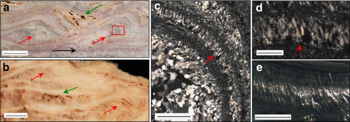
Scale bar measurements indicated. (a) Dresser terracettes (red arrows) with preserved primary porosity (green arrow) and a horizon containing Dresser stratiform geyserite (black arrow). Scale bar, 1 cm. Inset box of c. displays palisade fabric. (b) >1,800-year-old sinter terracettes (red arrows) with preserved primary porosity (green arrow) from a sinter buttress at Te Kopia, New Zealand. Scale bar, 1 cm. Micrographs in XPL of (c) Dresser palisade fabric oriented vertical to bedding (scale bar, 1 mm) and (d) close-up (scale bar, 250 μm). (e) Sinter with preserved palisade fabric, Te Kopia, New Zealand. Scale bar, 1 mm.
Somewhat thicker laminae in the Dresser geyserite (2–30 μm), compared to modern geyserite (500 nm–4 μm thick)13, may be attributed to diagenesis, whereby very fine laminae are destroyed in the transition from opal-A to microquartz19,24, as seen in Late Jurassic geyserite examples19. Significantly, the Dresser geyserite deposits are found directly overlying mineralized barite veins emblematic of subsurface hydrothermal fluid pathways, described in detail below (Figs 3 and 4 and Supplementary Fig. 1).
Figure 3: Large isopachous barite masses as mineralized hot spring pools.
figure3
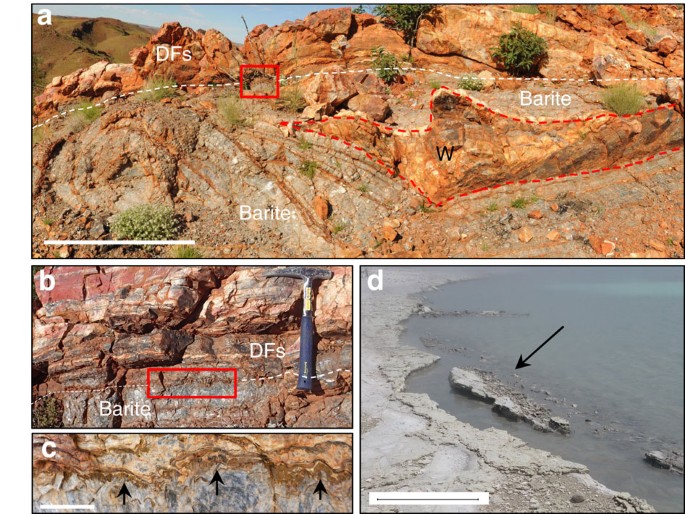
Scale bar measurements indicated. (a) Strongly curving isopachous barite veins envelop a chert wedge (W), overlain by sedimentary units (DFs). Scale bar, 1 m. Inset box of (b) isopachous layered barite underlies DFs that include stratiform geyserite. Inset box of (c) barite crystal tops growing upward into DFs (arrows). Scale bar, 2 cm. (d) Modern collapsing hot spring-pool lip edge, shoreline of Lake Rotokawa, Rotokawa geothermal area, New Zealand. Scale bar, 0.5 m.
Figure 4: Schematic model of active Dresser hot spring system and its fossilized mineralized remnants.
figure4
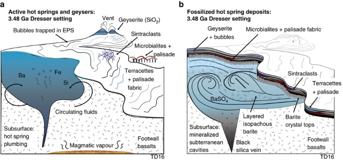
(a) Proximal to distal hot spring facies, with spring vent fed by subsurface hydrothermal fluids. (b) Preserved sequence of hot spring facies deposits, geographically patchy in nature, with spring vent infilled by late-stage crystallization of barite.
To distinguish the inferred geyserite deposits from other silica-replaced lithologies, such as microstromatolites13, or silicified sediments or travertines19, the composition of the siliceous light/dark laminae was examined in all Dresser samples using scanning electron microscopy-energy dispersive spectroscopy (SEM-EDS), Raman spectroscopy and X-ray diffraction (XRD) (mapping, point and bulk analyses; Supplementary Figs 3–7). SEM-EDS element maps show higher concentrations of Al and K within lighter bands, which contain singular and clustered lath-like flakes, 2–30 μm in size. Flakes are composed primarily of Si, O, Al and K with minor Na, Ba and Mg dispersed within a siliceous matrix (Si, O and very minor Al). XRD patterns indicate that the Al- and K-rich laths and flakey aggregates are composed of intermixed kaolinite (Al2Si2O5(OH)4) and illite (K,H3O)(Al,Mg,Fe)2(Si,Al)4O10[(OH)2,(H2O)]). SEM-EDS element maps display concentrations of Ti within darker laminae, revealed by SEM as very fine (2 μm) crystal grains composed dominantly of Ti and O, locally with trace amounts of K, Al and Na. However, these trace elements, as well as large silicon peaks, are considered to derive from the microquartz matrix (Si, O with minor K and Al±Na). At 20 kv, interaction/penetration of the beam is a minimum of 2–3 μm, whereas many of the Ti-grains are of a similar size (that is, 2 μm), or larger. Thus, it is unlikely that a discrete signal was acquired solely from the Ti-rich grains. Raman spectra showed major peaks at 143 and 638, indicative of anatase (TiO2), and a major peak at 464, indicative of quartz. Mineral confirmation XRD patterns indicate that dark laminae contain abundant, micron-scale, anatase crystals dispersed in a matrix of microquartz. Equigranular troughs between laminated botryoids contain relatively equal proportions of Ti, K and Al (Supplementary Fig. 7).
Both kaolinite+illite and anatase are documented alteration minerals in the upper level of geothermal fields, for example, at Sulphur Springs, New Mexico25, and the Soufriere Hills volcano, Montserrat26. Anatase has also been reported in Jurassic geyserites19. Kaolinite+illite is a diagnostic alteration mineral suite in shallow, advanced to intermediate argillic alteration (<120 epithermal="" high-sulfidation="" of="" sup="" systems="" zones="">24
, including that of the Dresser Formation, which displays steam-heated acid-sulfate kaolinite+illite alteration of underlying pillow basalts4. Precipitation of anatase is favoured in near neutral to alkaline pH27, which is consistent with formation of geyserite in modern low temperature (<100 alkali-chloride="" near="" neutral="" springs="" sup="" thermal="">19. The anatase is unlikely to be a retrograde alteration product of rutile (high-temperature polymorph of TiO2), as the latter is preserved in the higher temperature phyllic alteration zone within underlying basalts around the Dresser barite mine4, located <3 400="" a="" above="" anatase="" are="" aria-label="Reference 28" constraints="" containing="" data-test="citation-ref" data-track-action="reference anchor" data-track-label="link" data-track="click" dfc1="" from="" geyserite.="" href="https://www.nature.com/articles/ncomms15263#ref-CR28" id="ref-link-section-d52424e1134" in="" indicated="" inferred="" irreversibly="" km="" of="" preservation="" ref.="" rutile="" sites="" temperature="" title="Hanaor, D. A. & Sorrell, C. C. Review of the anatase to rutile phase transformation. J. Mater. Sci. 46, 855–874 (2011)." to="" transforms="" which="">28); presence of stable kaolinite+illite mineral assemblages, indicative of temperatures <120 and="" class="stix" cool="" data="" dfc1="" fluid="" from="" inclusion="" indicate="" relatively="" span="" which="">∼120 °C) water temperatures under low-confining pressures near the palaeosurface7.

Sinter terracettes

At locality 1S (Supplementary Fig. 1), stratiform geyserite is overlain by a 3 cm thick unit composed of siliceous, millimetre-thin laminae that form 1 cm diameter, low-amplitude (<2 a="" asymmetrical="" cm="" convex="" data-track-action="figure anchor" data-track-label="link" data-track="click" href="https://www.nature.com/articles/ncomms15263#Fig2" ridges="">Fig. 2a
). The laminae in these ridges are stacked into what superficially resemble climbing ripples, but differ in that thicker laminae appear on the down-current side. The convex laminae in cross-section resemble sinter terracettes (smaller-scale subsets within sinter terraces) from the mid- to distal-apron facies of hot springs, displaying the primary porosity and microtextures comparable with more recent, microbially derived examples19 (Fig. 2a,b).

Mineralized hot spring pools

New observations are presented here with respect to the formation mechanism of large barite masses found at the upper tips of black silica+barite veins, which are relevant in interpreting a terrestrial hot spring setting for DFc1. Subspherical (5–20 m diameter) masses of coarsely crystalline, isopachous, hydrothermal barite (+pyrite) occupy the uppermost parts of hydrothermal veins where they contact DFc1 sedimentary deposits. Previously, these barite masses were interpreted as baritized, diapiric gypsum bodies2, and later were inferred to be primary ‘barite mounds’ formed on the seafloor3. However, new observations suggest that at least some of these barite masses represent the mineralized remnants of terrestrial hot spring pools and associated shallow subsurface hydrothermal plumbing.
Significantly, large hydrothermal barite masses immediately underlie two of the geyserite localities described here, outcropping at the top of 10–20 m wide, 1 km deep silica+barite hydrothermal veins that cut their way up into the base of the finely layered sedimentary succession (Supplementary Fig. 1). The barite masses typically consist of multiple, thick, distinctly curved isopachous layers of coarsely crystalline barite (Fig. 3a) with crystals up to 10 cm long that consistently point upwards and outwards towards the edges of the masses. Typically, sets of barite crystals are separated by thin pyrite laminae from which sulfur isotopic values point to microbial disproportionation12. At locality 1S (Supplementary Fig. 1), a large hydrothermal barite mass displays distinct sets of isopachous barite layers with strongly curving geometries that envelop a wedge-shaped block of layered chert-barite derived from the overlying sedimentary succession (Fig. 3a). The uppermost barite layer has crystal tops that project into the base of the overlying sedimentary unit containing geyserite and siliceous sinter (Fig. 3b,c). Together, these observations suggest that barite mineralization developed beneath a collapsing, but semi-lithified sedimentary crust containing localized geyserite and siliceous sinter. This interpretation is supported by observations made 3 km north of the Dresser Mine, where a 10 m long × 1 m thick tilted panel of bedded chert+microbialites+vein barite, together with angular to rounded blocks of hydrothermal barite and chert, form a megabreccia (devoid of geyserite) that fills what was a large subspherical cavity with steeply dipping walls that cuts down through bedded chert (see for comparison Fig. 13, p. 215 of ref. 5). Formation of the megabreccia occurred during sediment accumulation, as demonstrated by bedded chert that overlies the cavity.
These barite masses occur at the uppermost tips of hydrothermal feeder veins along faults (that is, hydrothermal fluid conduits), and some are found immediately beneath overlying strata with known geyserite and siliceous sinter deposits. Therefore, these large enveloped and tilted sedimentary blocks are interpreted as collapsed hot spring terraces or pool margins such as those observed in modern geothermal areas (Fig. 3d).
The geometry and isopachous nature of these barite masses may be compared to fossilized travertine deposits of Lake Bogoria, Kenya, where isopachous carbonate layers systematically line and fill subterranean cavities of the former hot spring pools (see Fig. 4, p. 806 in ref. 29). While barite is not present in the Lake Bogoria example, the textures are equivalent. Although rare, terrestrial hot spring barite is known to precipitate alongside silica30, but no reported modern examples host the large quantities of barite found in DFc1. In summary, these data suggest that the isopachous barite masses represent the mineralized remnants of hot spring pools at the uppermost parts of the geothermal plumbing system (Fig. 4).

Biosignatures in Dresser hot spring deposits

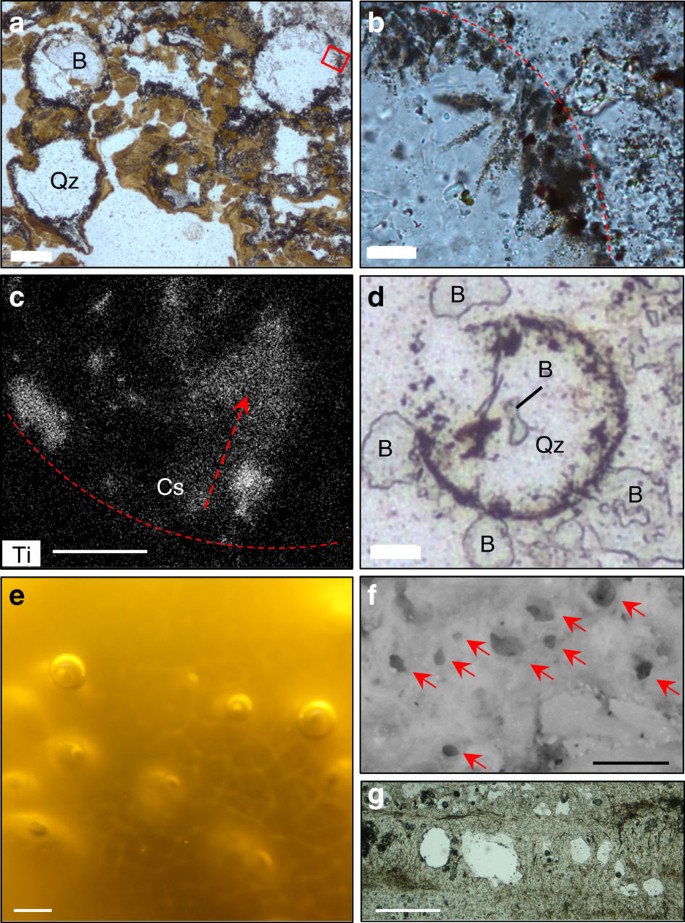
Horizons containing intergrown hydrothermal microquartz and barite interspersed with Fe-oxyhydroxides (the latter a product of Tertiary weathering of primary pyrite) occur between sets of the fine light/dark siliceous microlaminae within the Dresser columnar–botryoidal geyserite at locality 16N (Fig. 1a and Supplementary Fig. 8). Contained within these horizons are numerous circular to sub-circular structures, 200 μm in diameter, filled by microquartz and barite, but lined with fine-grained anatase and internal anatase crystal splays that fan inwards from the margins towards the centre of the structures (Fig. 5a–c). The infilling microquartz and barite cuts across the anatase-lined walls of the structures, indicative of their early formation during DFc1 hydrothermal activity (Fig. 5a,d).
Figure 5: Inferred gas bubbles.
figure5
Scale bar measurements indicated. Micrographs in PPL (a,b). (a) Spherical to subspherical inferred bubbles filled with quartz (Qz) and barite (B). Scale bar, 90 μm. Inset box of (b) inward radiating anatase crystal splays from bubble rim (dashed line). Scale bar, 22 μm. (c) SEM-EDS element map data of Ti concentration in crystal splay (Cs) along bubble rim (dashed line). Scale bar, 9 μm. (d) Quartz (Qz) and barite (B) cutting bubble wall. Scale bar, 22 μm. (e) Oxygen bubbles in modern EPS of a mid-apron cyanobacterial mat, Orakei Korako, New Zealand. Scale bar, 2 mm. (f) Spherical to subspherical fossil bubbles (arrows) preserved in wavy laminated sinter, representing a silicified microstromatolite+EPS typical of mid-temperature sinter apron pools from the recently extinct Beowawe hot spring, Nevada. Scale bar, 200 μm. (g) Microbial palisade fabric with spherical structures, representing silicified gas bubbles in 15 Ka sinter, Tahunaatara, Taupo Volcanic Zone, New Zealand. Scale bar, 250 μm.
These circular structures are discounted as fluid inclusions based on their larger size (20 × larger than typical fluid inclusions at 10 μm diameter) and their occurrence in a polymineralic matrix7. Rather, the structures are interpreted as gas bubbles31. Many gas bubbles, both biogenic and abiogenic, are subject to deformation caused by their host lithology31,32, yet most Dresser bubbles retain a circular outline with only a few showing collapsed margins. Their excellent preservation suggests growth within, and perhaps partial passage through, an elastic medium that also trapped them before they reached the surface and burst. The only known medium in hot spring settings with such properties is exopolymeric substance (EPS)31 derived from microbial mats/biofilms that flourish on the mid-aprons of modern sinter terraces (Fig. 5e). Entrapment of bubbles in EPS would allow for retention of the bubble structure while allowing for internal, inward radiating crystallization of anatase during hydrothermal alteration of the biofilm.
Alternately, bubbles observed floating on the surface of hot spring pools from Mammoth Hot Springs at Yellowstone National Park were reported as having preserved their shape via calcite crystallization33. However, such crystals radiate outward from bubble surfaces and thus contrast with the internal, inward radiating anatase crystals in the Dresser bubbles. Rather, entrapment in EPS would retain the bubble structure while allowing internal, inward radiating crystallization.
Therefore, regardless of whether the Dresser bubbles were derived from degassing of thermal fluids or represent metabolic gas derivatives, preservation likely occurred almost immediately, through entrapment in microbial EPS. Bubbles are commonly preserved in microbial sinter within mid-apron hot spring facies via trapping of microbial exudate (for example, oxygen)15,16. In modern examples, bubbles become silicified along with the microbial mat and either become infilled with sinter/microbial filaments or remain open16. Those forming in channels become flattened and appear almond shaped in cross-section17, whereas bubbles formed in quiet, mid-temperature (45–55 °C) apron pools may preserve spherical shapes16 (Fig. 5f,g). The stratigraphic association of geyserite and horizons with bubbles in EPS can be explained as a function of Walther’s Law, owing to laterally shifting discharge conditions or intermittent decreases in spring outflow temperatures, as vent geyserite can be found interbedded with mid- and low-temperature sinter apron fabrics34.
In addition, within the unit of sinter terracettes, some thin laminae display vertically aligned quartz crystals (230 μm high) that wrap around the curved hinge of the convex ridges (Fig. 2c,d). Epithermal vein textures are discounted as there is no evidence of cross-cutting veins. Similarly, the vertically aligned fabric extends for many centimetres along bedding, whereas veins typically display quartz infill of an open cavity (Supplementary Fig. 9a). Shearing textures are also discounted since these would display quartz crystals aligned all in the same direction, rather than fanning around convex boundaries as is observed in the Dresser fabric (Supplementary Fig. 9b,c). Association with hot spring geyserite and the observation that the inferred palisade fabric is situated within interpreted sinter terracettes provide contextual support for formation in a hot spring setting. Therefore, these vertically aligned quartz crystals are suggested as analogous to recrystallized microbial palisade fabric formed through silicification of microbial filaments oriented perpendicular to bedding surfaces on mid- to distal-apron hot spring terraces19 (Fig. 2e and Supplementary Fig. 9d).
Finally, a link between hot spring deposits and macroscopic stromatolites is drawn from geyserite rip-up clasts found interbedded with elongate domical and conical stromatolitic laminates (locality 24S: Supplementary Fig. 1) composed of ferruginized laminae (altered pyrite based on drill core comparisons6), and draped by ribbons and shards of felsic volcanic ash mixed with sand grains. Onlapping by these tuffaceous sediments and the irregular internal laminae with faint palimpsest microfabrics within these stromatolites are consistent with a microbial origin (Supplementary Fig. 10).

Discussion

Textural similarity of the black-and-white laminated, laminar to botryoidal siliceous Dresser deposits with modern and Phanerozoic geyserite, combined with mineralogy consistent with geothermal settings, provide support for the previously unrecognized presence of geyserite in the Dresser Formation. Geyserite is known to precipitate from hot (>75–100 °C), silica-rich, near-neutral pH, alkali-chloride fluids ejected from boiling pools and geysers on exposed land surfaces18,19. A link between these surface fluids and the steam-heated acid sulfate alteration at the Dresser Mine4 is provided by comparative mineralogy and studies of active geothermal systems. Steam-heated acid sulfate alteration forming kaolinite+illite mineral assemblages occurs in the late stages of an evolving high-sulfidation system24. In such a system, initial alkali-chloride geothermal springs subsequently develop into an acid steam-dominated system, commonly due to a drop in the water table under fluctuating, or waning, thermal activity24. In addition to geyserite, the discovery of siliceous sinter with terracettes and the mineralized remnants of hot spring pools collectively indicate a period of exposed land surface with terrestrial hot springs during Dresser deposition.
A hot spring setting is supported by the abrupt lateral and temporal facies changes observed over short distances (to the millimetre scale), including the patchy spatial distribution of geyserite, sinter and fluvial deposits. Inferred fluvial features include shallow channelized pebble to cobble conglomerate, distinctive edgewise conglomerate and cross-rippled sedimentary rocks. Such rapid changes are consistent with the ‘tremendous variability observed in all siliceous hot springs’34. Variability is controlled by fluctuations in hot spring discharge, which influence temperature, flow rate, sinter facies and microbial growth34. This lateral and temporal variability contrasts markedly with Archaean marine or lacustrine deposits that are characterized by zoned stromatolite morphologies that typically show some degree of lateral and/or temporal continuity over tens, to hundreds, and even thousands of metres13,35. Additionally, neither marine nor lacustrine settings can account for the preservation of geyserite, sinter terracettes or the mineralized remnants of hot spring pools.
Importantly, all recognized Dresser hot spring facies contain, or are spatially associated with, a suite of newly identified inferred biosignatures, including iron-rich domical and conical stromatolites, microbial palisade fabric within sinter terracettes and silicified bubbles in microbial EPS on sinter apron deposits. These observations suggest that early life in the Dresser Formation thrived off the chemical energy in hot springs.
In conclusion, newly discovered terrestrial hot spring facies in the ca. 3.5 Ga Dresser Formation contain a range of highly distinctive and varied textural biosignatures, providing direct evidence that at least some of Earth’s earliest life thrived on land, in hot springs (Fig. 4). The Dresser Formation terrestrial hot spring facies include geyserite, siliceous sinter terracettes and the mineralized remnants of hot spring pools. These findings extend the geological record of inhabited terrestrial hot springs by 3 billion years, the occurrence of an exposed land surface by up to 130 million years36,37 and evidence of life on land by 580 million years21.
This result is significant in that it further constrains our understanding of the evolution of early life on Earth, as well as offers astrobiological implications in the search for potential fossil life on Mars. The Dresser Formation shares a similar age to older portions of the Martian crust and provides the closest comparison to geological processes likely occurring on Mars at that time38. The similarity of the Dresser deposits to modern hot springs shows that ancient hot spring processes on Earth were not so different from today. This lends weight to the use of modern and Phanerozoic terrestrial analogues in exploring for life in fossil Martian hot springs19,39, and demonstrates the exceptional preservation potential of these very ancient fossil-bearing hot spring deposits here on Earth.

Methods

Mapping

To assess possible associations between stromatolitic surface features and subsurface circulating hydrothermal fluids preserved in extensive vein networks, detailed mapping was undertaken to constrain geological context in a 7 km strike-length study area centred on the Dresser barite mine; 47 stratigraphic sections were measured. Voluminous amounts of barite were deposited as a result of extensive hydrothermal fluid circulation and therefore sedimentary units relatively proximal to these fluid conduits were chosen for in-depth investigation. Aerial photographs and geological maps of the area were used to navigate the terrain. GPS coordinates were collected from each locality for relocation.

Light microscopy

Optical light microscopy was conducted at the University of New South Wales on a Leica DM2500 with LAS 3.6 software to acquire photomicrographs.

Raman spectroscopy

Raman spectroscopy was measured at the Mark Wainwright Analytical Centre, UNSW using a Reni Shaw inVia Raman spectrometer using an argon ion laser providing excitation at 514 nm and fitted with an automated XYZ microscope stage. A 1 × 1 mm region of laminations on the petrographic thin section was selected using a × 50 objective and a series of measurements from 115–2,000 cm−1 were collected at intervals of 25 μm. Laser spot sizes of 1 μm were estimated using this objective. Images were prepared using the Renishaw WiRE software by displaying the height of one major peak for each of the mineral phases of interest. Only two major mineral phases, quartz and anatase, were observed in the measured region. The spectra of quartz and anatase were examined, and one peak was selected from each spectrum that was unique to that mineral, with no overlap from other peaks. The intensity of quartz (red) was displayed using the peak at 484.8 cm−1. The intensity of anatase (green) was displayed using the peak at 637.4 cm−1. Images were blended, then superimposed on an incident white light image of the sample taken using a Leica M165C microscope.

SEM-EDS

Standard 30 μm thick petrographic thin sections were prepared with an evaporative carbon coating at the electron microscopy unit of the Mark Wainwright Analytical Centre at the University of New South Wales Australia. Samples were examined using a Hitachi S-3400N SEM operating at 20 kV and fitted with a Bruker SDD-EDS XFlash 6–30 detector.

XRD

In situ XRD analysis of the putative botryoidal geyserite was carried out by a Bruker D8 TXS with a basic parameter operating at 45 kV and 100 mA. The scan included 5–85 degrees (2Q). Micro-diffraction optics were used with a beam size of 0.5 mm.

Data availability

The authors declare that all data supporting the findings of this study are available within the paper (and its Supplementary Information files).

Additional information

How to cite this article: Djokic, T. et al. Earliest signs of life on land preserved in ca. 3.5 Ga hot spring deposits. Nat. Commun. 8, 15263 doi: 10.1038/ncomms15263 (2017).
Publisher’s note: Springer Nature remains neutral with regard to jurisdictional claims in published maps and institutional affiliations.

Change history

  • 16 August 2017

    A correction has been published and is appended to both the HTML and PDF versions of this paper. The error has not been fixed in the paper.

References

  1. 1
    Walter, M. R., Buick, R. & Dunlop, J. S. R. Stromatolites 3,400–3,500 Myr old from the North Pole area, Western Australia. Nature 248, 443–445 (1980).
  2. 2
    Buick, R. & Dunlop, J. S. R. Evaporitic sediments of early Archaean age from the Warrawoona Group, North Pole, Western Australia. Sedimentology 37, 247–277 (1990).
  3. 3
    Nijman, W., de Bruijne, K. C. H. & Valkering, M. E. Growth fault control of early Archaean cherts, barite mounds and chert-barite veins, North Pole Dome, Eastern Pilbara, Western Australia. Precambrian Res. 88, 25–52 (1998).
  4. 4
    Van Kranendonk, M. J. & Pirajno, F. Geochemistry of metabasalts and hydrothermal alteration zones associated with c. 3.45 Ga chert and barite deposits: implications for the geological setting of the Warrawoona Group, Pilbara Craton, Australia. Geochemistry 4, 253–278 (2004).
  5. 5
    Van Kranendonk, M. J. Volcanic degassing, hydrothermal circulation and the flourishing of early life on Earth: a review of the evidence from c. 3490-3240 Ma rocks of the Pilbara Supergroup, Pilbara Craton, Western Australia. Earth Sci. Rev. 74, 197–240 (2006).
  6. 6
    Van Kranendonk, M. J., Philippot, P., Lepot, K., Bodorkos, S. & Pirajno, F. Geological setting of Earth's oldest fossils in the ca. 3.5 Ga Dresser Formation, Pilbara Craton, Western Australia. Precambrian Res. 167, 93–124 (2008).
  7. 7
    Harris, A. C. et al. Early Archean hot springs above epithermal veins, North Pole, Western Australia: new insights from fluid inclusion microanalysis. Econ. Geol. 104, 793–814 (2009).
  8. 8
    Ueno, Y., Yoshioka, H., Maruyama, S. & Isozaki, Y. Carbon isotopes and petrography of kerogens in 3.5-Ga hydrothermal silica dikes in the North Pole area, Western Australia. Geochim. Cosmochim. Acta 68, 573–589 (2004).
  9. 9
    Morag, N. et al. Microstructure-specific carbon isotopic signatures of organic matter from 3.5 Ga cherts of the Pilbara Craton support a biologic origin. Precambrian Res. 275, 429–449 (2016).
  10. 10
    Van Kranendonk, M. J. in Advances In Stromatolite Geobiology (ed. Reitner, J.) 537–554 (Springer, 2011).
  11. 11
    Glikson, M. et al. Microbial remains in some earliest Earth rocks: comparison with a potential modern analogue. Precambrian Res. 164, 187–200 (2008).
  12. 12
    Shen, Y., Farquhar, J., Masterson, A., Kaufman, A. J. & Buick, R. Evaluating the role of microbial sulfate reduction in the early Archean using quadruple isotope systematics. Earth Planet. Sci. Lett. 279, 383–391 (2009).
  13. 13
    Walter, M. R. in Stromatolites (ed. Walter, M. R.) 87–112 (Elsevier, 1976).
  14. 14
    Cady, S. L. & Farmer, J. D. in Evolution Of Hydrothermal Ecosystems On Earth (And Mars?). Ciba foundation symposium 202 (eds Bock, G. R. & Goode, G. A.) 150–173 (John Wiley, 1996).
  15. 15
    Hinman, N. W. & Walter, M. R. Textural preservation in siliceous hot spring deposits during early diagenesis: examples from Yellowstone National Park and Nevada, USA. J. Sediment. Res. 75, 200–215 (2005).
  16. 16
    Handley, K. M. & Campbell, K. A. in Stromatolites: interaction of Microbes with Sediments (eds Tewari, V. & Seckbach, J.) 359–381 (Springer, 2011).
  17. 17
    Lynne, B. Y. Mapping vent to distal-apron hot spring paleo-flow pathways using siliceous sinter architecture. Geothermics 43, 3–24 (2012).
  18. 18
    White, D. E., Thompson, G. A. & Sandberg, C. H. Rocks, structure, and geologic history of Steamboat Springs thermal area, Washoe County, Nevada. U.S. Geol. Surv. Prof. Paper 458-B (1964).
  19. 19
    Campbell, K. A. et al. Geyserite in hot-spring siliceous sinter: window on Earth's hottest terrestrial (paleo) environment and its extreme life. Earth Sci. Rev. 148, 44–64 (2015).
  20. 20
    Konhauser, K. O., Phoenix, V. R., Bottrell, S. H., Adams, D. G. & Head, I. M. Microbial–silica interactions in Icelandic hot spring sinter: possible analogues for some Precambrian siliceous stromatolites. Sedimentology 48, 415–433 (2001).
  21. 21
    Beraldi-Campesi, H. Early life on land and the first terrestrial ecosystems. Ecol. Process. 2, 1 (2013).
  22. 22
    Dong, G., Morrison, G. & Jaireth, S. Quartz textures in epithermal veins, Queensland; classification, origin and implication. Econ. Geol. 90, 1841–1856 (1995).
  23. 23
    Jones, B. & Renaut, R. W. Hot spring and geyser sinters: the integrated product of precipitation, replacement, and deposition. Can. J. Earth. Sci. 40, 1549–1569 (2003).
  24. 24
    Sillitoe, R. H. Epithermal paleosurfaces. Miner. Deposita 50, 767–793 (2015).
  25. 25
    Charles, R., Buden, R. & Goff, F. An interpretation of the alteration assemblages at Sulphur Springs, Valles caldera, New Mexico. J. Geophys. Res. 91, 1887–1898 (1986).
  26. 26
    Boudon, G., Villemant, B., Komorowski, J. C., Ildefonse, P. & Semet, M. P. The hydrothermal system at Soufriere Hills Volcano, Montserrat (West Indies): characterization and role in the on‐going eruption. Geophys. Res. Lett. 25, 3693–3696 (1998).
  27. 27
    Kinsinger, N. M., Wong, A., Li, D., Villalobos, F. & Kisailus, D. Nucleation and crystal growth of nanocrystalline anatase and rutile phase TiO2 from a water-soluble precursor. Cryst. Growth Des. 10, 5254–5261 (2010).
  28. 28
    Hanaor, D. A. & Sorrell, C. C. Review of the anatase to rutile phase transformation. J. Mater. Sci. 46, 855–874 (2011).
  29. 29
    Renaut, R. W. & Jones, B. Controls on aragonite and calcite precipitation in hot spring travertines at Chemurkeu, Lake Bogoria, Kenya. Can. J. Earth. Sci. 34, 801–818 (1997).
  30. 30
    Bonny, S. M., Jones, B. & Rankey, G. Petrography and textural development of inorganic and biogenic lithotypes in a relict barite tufa deposit at Flybye Springs, NT, Canada. Sedimentology 55, 275–303 (2008).
  31. 31
    Flugel, E. Microfacies of Carbonate Rocks: analysis, Interpretation and Application Springer (2004).
  32. 32
    Wilmeth, D. T. et al. Punctuated growth of microbial cones within early Cambrian oncoids, Bayan Gol Formation, Western Mongolia. Palaios 30, 836–845 (2015).
  33. 33
    Chafetz, H. S. & Folk, R. L. Travertines: depositional morphology and the bacterially constructed constituents. J. Sediment. Petrol. 54, 289–316 (1984).
  34. 34
    Guidry, S. A. & Chafetz, H. S. Anatomy of siliceous hot springs: examples from Yellowstone National Park, Wyoming, USA. Sediment. Geol. 157, 71–106 (2003).
  35. 35
    Awramik, S. M. & Buchheim, H. P. A giant, late Archean lake system: the Meentheena Member (Tumbiana Formation; Fortescue Group), Western Australia. Precambrian Res. 174, 215–240 (2009).
  36. 36
    Buick, R. et al. Record of emergent continental crust 3.5 billion years ago in the Pilbara Craton of Australia. Nature 375, 574–577 (1995).
  37. 37
    Van Kranendonk, M. J., Hugh Smithies, R., Hickman, A. H. & Champion, D. Review: secular tectonic evolution of Archean continental crust: interplay between horizontal and vertical processes in the formation of the Pilbara Craton, Australia. Terra Nova 19, 1–38 (2007).
  38. 38
    Walter, M. & Des Marais, D. J. Preservation of biological information in thermal spring deposits: developing a strategy for the search for fossil life on Mars. Icarus 101, 129–143 (1993).
  39. 39
    Ruff, S. W. & Farmer, J. D. Silica deposits on Mars with features resembling hot spring biosignatures at El Tatio in Chile. Nat. Commun. 7, 13554 (2016).
Download references

Acknowledgements

Many thanks to: J. Reinter for discussion and assistance with Raman spectroscopic data; C. Marjo for assistance with Raman spectroscopic data; K. Privat for assistance with SEM-EDS data and the electron microscope unit, UNSW. Research support provided by the Australian Centre for Astrobiology and School of Biological, Earth and Environmental Sciences at the University of New South Wales, the Sloan Foundation and the ARC Centre for excellence Core to Crust Fluid Systems. Phanerozoic hot spring comparative studies were supported by funding to K.A.C. from the New Zealand government (RSNZ Marsden Fund and Ministry of Business, Innovation and Employment) and the National Geographic Society. Gigapan image generated by Ken Williford and the abcLab, Jet Propulsion Laboratory, California Institute of Technology. Kind hospitality in the field was provided by Faye and Geoff Myers, and Haoma Mining.

Author information

The methodology was conceived and designed by T.D. and M.J.V.K. Geological mapping was carried out by T.D. and M.J.V.K. Petrographic analyses were carried out by T.D., M.J.V.K., K.A.C. and M.R.W. SEM-EDS data were acquired and interpreted by T.D. and M.J.V.K. XRD analysis spectra were acquired by C.R.W. All authors contributed to discussion, interpretation and writing.
Correspondence to Tara Djokic.

Ethics declarations

Competing interests

The authors declare no competing financial interests.

Supplementary information

Supplementary Information

Supplementary Figures (PDF 20800 kb)

Peer Review File (PDF 350 kb)

Rights and permissions

This work is licensed under a Creative Commons Attribution 4.0 International License. The images or other third party material in this article are included in the article’s Creative Commons license, unless indicated otherwise in the credit line; if the material is not included under the Creative Commons license, users will need to obtain permission from the license holder to reproduce the material. To view a copy of this license, visit http://creativecommons.org/licenses/by/4.0/
Reprints and Permissions

About this article

Verify currency and authenticity via CrossMark

Cite this article

Djokic, T., Van Kranendonk, M., Campbell, K. et al. Earliest signs of life on land preserved in ca. 3.5 Ga hot spring deposits. Nat Commun 8, 15263 (2017). https://doi.org/10.1038/ncomms15263