Fossil soft tissue structure
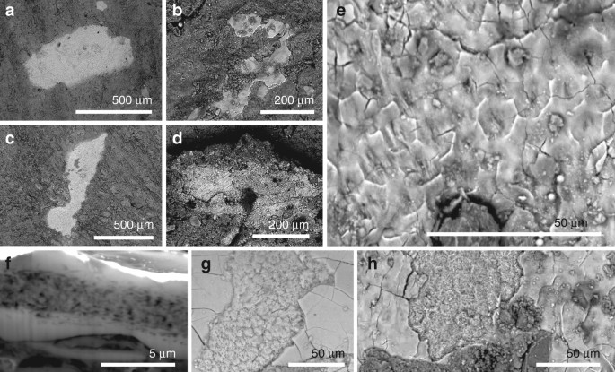
Small patches of tissue (0.01–0.4 mm
2; Fig.
1a–d and Supplementary Figs.
2–
6) are closely associated with fossil feathers (i.e., usually within 500 µm of carbonaceous feather residues, Supplementary Fig.
2e, g, j, k, o, s, t).
The patches are definitively of fossil tissue, and do not reflect
surface contamination with modern material during sample preparation, as
they are preserved in calcium phosphate (see 'Taphonomy', below);
further, several samples show margins that are overlapped, in part, by
the surrounding matrix. The tissues have not, therefore, simply adhered
to the sample surface as a result of contamination from airborne
particles in the laboratory.
The tissue patches are typically 3–6 µm thick and planar (Fig.
1a–e).
Transverse sections and fractured surfaces show an inner fibrous layer
(1.0–1.2 µm thick) between two thinner structureless layers (0.2–0.5 µm
thick) (Fig.
1f–h).
The external surface of the structureless layer is smooth and can show
a subtle polygonal texture defined by polygons 10–15 µm wide (Fig.
1e, h).
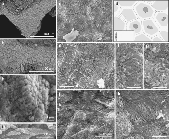
The fibrous layer also shows polygons (Figs.
1f, h and
2a–e, and Supplementary Fig.
6) that contain arrays of densely packed fibres 0.1–0.5 µm wide (Fig.
2f–i and Supplementary Fig.
5f). Well-preserved fibres show helicoidal twisting (Fig.
2h, i).
Fibres in marginal parts of each polygon are 0.1–0.3 µm wide and
oriented parallel to the tissue surface; those in the interior of each
polygon are 0.3–0.5 µm wide and are usually perpendicular to the tissue
surface (Fig.
2b, h and Supplementary Fig.
S6d).
In the marginal 1–2 µm of each polygon, the fibres are usually
orthogonal to the lateral polygon margin and terminate at, or bridge the
junction between, adjacent polygons (Fig.
2f, g and Supplementary Fig.
6e).
The polygons are usually equidimensional but are locally elongated and
mutually aligned, where the thick fibres in each polygon are
sub-parallel to the tissue surface and the thin fibres, parallel to the
polygon margin (Fig.
2j, k and Supplementary Fig.
6g–l). Some polygons show a central depression (Fig.
2c–e and Supplementary Fig.
6a–c) in which the thick fibres can envelop a globular structure 1–2 µm wide (Fig.
2e).
Fossil corneocytes
The
texture of these fossil tissues differs from that of conchostracan
shells and fish scales from the host sediment, the shell of modern
Mytilus, modern and fossil feather rachis and modern reptile epidermis (Supplementary Fig.
7a–n). The elongate geometry of some polygons (Fig.
2j, k and Supplementary Fig.
6g, l)
implies elastic deformation of a non-biomineralized tissue due to
mechanical stress. On the basis of their size, geometry and internal
structure, the polygonal structures are interpreted as corneocytes
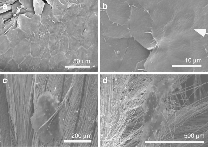
(epidermal keratinocytes). In modern amniotes, these are
polyhedral-flattened cells (1–3 µm × ca. 15 µm) filled with keratin
tonofibrils, lipids and matrix proteins
18,19,20 (Fig.
3a, b and Supplementary Figs
2u–x,
8,
9).
The outer structureless layer of the fossil material corresponds to the
cell margin; it is thicker than the original biological template, i.e.,
the corneous cell envelope and/or cell membrane, but this is not
unexpected, reflecting diagenetic overgrowth by calcium phosphate (see
'Taphonomy'). The fibres in the fossil corneocytes are identified as
mineralised tonofibrils: straight, unbranching bundles of supercoiled
α-keratin fibrils 0.25–1 µm wide
18,21 that are the main component of the corneocyte cytoskeleton
22 and are enveloped by amorphous cytoskeletal proteins
22. In the fossils, the thin tonofibrils often abut those of the adjacent cell (Fig.
2g and Supplementary Fig.
6e), but locally can bridge the boundary between adjacent cells (Fig.
2f). The latter recalls desmosomes, regions of strong intercellular attachment between modern corneocytes
23. The central globular structures within the fossil corneocytes resemble dead cell nuclei
24, as in corneocytes of extant birds (but not extant reptiles and mammals)
24 (Supplementary Fig.
8). The position of these pycnotic nuclei is often indicated by depressions in the corneocyte surface in extant birds
24 (Fig.
3b); some fossil cells show similar depressions (Fig.
2c and Supplementary Fig.
6a–c).
Taphonomy
Keratin
is a relatively recalcitrant biomolecule due to its heavily
cross-linked paracrystalline structure and hydrophobic nonpolar
character
23.
Replication of the fossil corneocytes in calcium phosphate is thus
somewhat unexpected as this process usually requires steep geochemical
gradients characteristic of early decay
25 and usually applies to decay-prone tissues, such as muscle
26 and digestive tissues
27.
Recalcitrant tissues such as dermal collagen can, however, be
replicated in calcium phosphate where they contain an inherent source of
calcium and, in particular, phosphate ions that are liberated during
decay
28.
Corneocytes contain sources of both of these ions. During terminal
differentiation, intracellular concentrations of calcium increase
29 and α-keratin chains are extensively phosphorylated
23. Further, corneocyte lipid granules
30 are rich in phosphorus and phosphate
31.
These chemical moieties would be released during degradation of the
granules and would precipitate on the remaining organic substrate, i.e.,
the tonofibrils.
In extant mammals, densely packed arrays of tonofibrils require abundant interkeratin matrix proteins for stability
32.
These proteins, however, are not evident in the fossils. This is not
unexpected, as the proteins are rare in extant avian corneocytes
33 and, critically, occur as dispersed monomers
34
and would have a lower preservation potential than the highly
cross-linked and polymerised keratin bundles of the tonofibrils. The
outer structureless layer of the fossil corneocytes is thicker than the
likely biological template(s), i.e., the corneous cell envelope (a layer
of lipids, keratin and other proteins up to 100 nm thick that replaces
the cell membrane during terminal differentiation
34)
and/or cell membrane. This may reflect a local microenvironment
conducive to precipitation of calcium phosphate: during terminal
differentiation, granules of keratohyalin, an extensively phosphorylated
protein
35 with a high affinity for calcium ions
36, accumulate at the periphery of the developing corneocytes
37.
The thickness of the outer solid layer of calcium phosphate in the
fossils, plus the gradual transition from this to the inner fibrous
layer, suggests that precipitation of phosphate proceeded from the
margins towards the interior of the corneocytes. In this scenario,
phosphate availability in the marginal zones of the cells would have
exceeded that required to replicate the tonofibrils. The additional
phosphate would have precipitated as calcium phosphate in the
interstitial spaces between the tonofibrils, progressing inwards from
the inner face of the cell margin.
Skin shedding in feathered dinosaurs and early birds
In
extant amniotes, the epidermal cornified layer is typically 5–20 cells
thick (but thickness varies among species and location on the body
38). The patches of fossil corneocytes, however, are one cell thick (Fig.
1f and Supplementary Figs.
5c,
10).
This, plus the consistent small size (<400 a="" and="" continuous="" fidelity="" high="" i="" in="" inconsistent="" is="" m="" minority="" nbsp="" of="" patches="" preservation="" remarkably="" selective="" sheet="" situ="" the="" tissue.="" with="">n
 46.7k
46.7k
 30.2k
30.2k
 11.7k
11.7k
 6.8k
6.8k