A 'ameba de fogo' sobrevive em condições mais quentes do que qualquer outra célula complexa.
O organismo unicelular consegue crescer a 63 °C, um recorde para a vida eucariótica.
Por
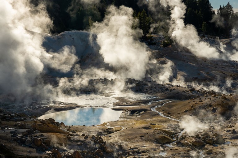
Hot springs at at Lassen Volcanic National Park in California, where the heat-tolerant amoeba was discovered.Credit: Kelly Vandellen/iStock via Getty
This
cell likes it hot. A tiny, single-celled amoeba that can thrive at
temperatures that kill all other known complex life — organisms whose
cells contain a nucleus and internal structures — has been found.
A
descoberta questiona a noção de que essa vida 'eucariótica' — que
inclui todos os animais e plantas — não seja adequada ao tipo de
condições extremas que podem ser toleradas por bactérias e outros
organismos que não possuem núcleo celular.
“Precisamos repensar
significativamente o que é possível para uma célula eucariótica”, afirma
Angela Oliverio, microbiologista da Universidade de Syracuse, em Nova
York. O trabalho, que ainda não foi revisado por pares, foi descrito em
um artigo pré-publicado. 1 Publicado em 24 de novembro.
Oliverio
e sua colega microbiologista de Syracuse, Beryl Rappaport, faziam parte
da equipe que descobriu o organismo no Parque Nacional Vulcânico
Lassen, na cordilheira das Cascatas, no norte da Califórnia. Eles o
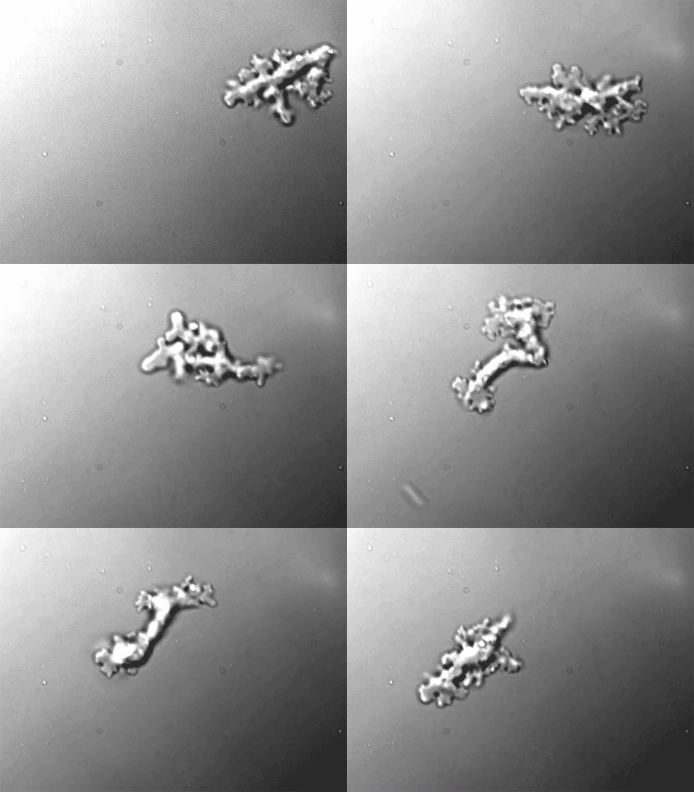
batizaram de Incendiamoeba cascadensis , que significa "ameba de fogo das cascatas".
Incendiamoeba cascadensis can thrive at temperatures that would kill other complex cells.Credit: Image taken by Natalie Petek-Seoane from the preprint by H. Beryl Rappaport et al./bioRxiv
O parque é famoso por seus lagos ácidos borbulhantes e piscinas geotérmicas incandescentes, mas a espécie I. cascadensis
provém de um "riacho quente" com pH neutro. "É a formação geotérmica
mais desinteressante que você encontrará em Lassen", diz Rappaport.
Amostras
de água do riacho pareciam desprovidas de vida sob um microscópio, mas
depois de cultivá-las com nutrientes, os pesquisadores observaram a
ameba crescendo a 57 °C, dentro da faixa de temperatura do riacho. Os
cientistas aumentaram a temperatura gradualmente, ultrapassando o
recorde anterior para eucariotos de 60 °C. A I. cascadensis
ainda era capaz de se dividir a 63 °C e continuava se movimentando a 64
°C. Mesmo a 70 °C, as células conseguiam formar "cistos" dormentes que
eram capazes de se reativar em temperaturas mais baixas.
As bactérias e arqueas mais resistentes conseguem suportar temperaturas muito mais elevadas: a arquea Methanopyrus kandleri
detém o recorde para qualquer forma de vida, com 122 °C. Algumas
espécies de fungos e algas vermelhas detinham o recorde anterior para
eucariotos; o limite superior para células humanas e de outros mamíferos
parece estar em torno de 43 °C.
A descoberta de I. cascadensis
ressalta o que pode ser ganho ao vasculhar o planeta em busca de novos
organismos, diz Julia Van Etten, bióloga evolucionista da Universidade
de Maryland, em College Park. "Este grupo descobriu um organismo que faz
algo que não sabíamos que os eucariotos eram capazes de fazer. Então, o
que mais existe por aí?"
Os pesquisadores têm dado pouca atenção
aos eucariotos que conseguem suportar condições extremas, acrescenta
Oliverio, que poderiam fornecer informações importantes para a busca de
vida além da Terra, bem como para a biotecnologia. "Nós analisamos
apenas um tipo de vida", diz ela. "Talvez tenhamos tido muita sorte e
não haja mais nada lá fora, mas realmente não acreditamos que seja esse o
caso."
doi: https://doi.org/10.1038/d41586-025-03933-5
Atualizações e correções
Correção de 02 de dezembro de 2025 : Em uma versão anterior deste artigo, o nome Incendiamoeba cascadensis estava grafado incorretamente. A grafia foi corrigida.
segunda-feira, 26 de dezembro de 2022
Estranho micróbio com tentáculos pode se assemelhar ao ancestral da vida complexa
Imagens internas detalhadas do segundo micróbio Asgard revelam componentes semelhantes às células vegetais e animais
Este micróbio recém-cultivado, apelidado de Lokiarchaeum ossiferum , pode se assemelhar a antigos precursores das células complexas que compõem plantas e animais. Thiago Rodrigues-Oliveira/Universidade de Viena
Ao cultivar um
micróbio com tentáculos incomum no laboratório, os microbiologistas
podem ter dado um grande passo para resolver os primeiros ramos da
árvore da vida e desvendar um de seus grandes mistérios: como as células
complexas que compõem o corpo humano - e todas as plantas, animais e
muitos organismos unicelulares - surgiram primeiro. Esses micróbios,
chamados Asgard archaea, já foram cultivados - uma vez -, mas o avanço
relatado hoje na Nature marca a primeira vez que eles foram cultivados em concentrações altas o suficiente para estudar suas entranhas em detalhes .
As imagens de microscopia
eletrônica resultantes revelam estruturas internas complexas sugestivas
daquelas em nossas próprias células, acrescentando suporte à ideia ainda
controversa de que micróbios antigos semelhantes a Asgard podem ter
sido os principais ancestrais de células complexas. O micróbio, da lama
de 15 centímetros de profundidade em um canal em um estuário na
Eslovênia, possui um citoesqueleto complexo feito da proteína actina,
sugerindo que essa estrutura surgiu em archaea antes de se tornar parte
integrante de células vegetais e animais. Essas descobertas se somam a
trabalhos recentes mostrando que as arqueas de Asgard possuem genes que
antes se pensava existirem apenas em organismos mais complexos – outra
indicação de que eles podem ser um importante precursor evolutivo.
"Este papel é lindo", diz Buzz
Baum, biólogo celular evolutivo do Laboratório de Biologia Molecular do
Conselho de Pesquisa Médica. “As imagens são impressionantes.” Baum
suspeita que muitos pesquisadores vão querer estudar os micróbios
recém-cultivados.
Considerado um terceiro
domínio da vida pela maioria dos cientistas, os archaea são distintos
das bactérias e dos eucariotos, o ramo evolutivo que inclui os humanos.
Ainda assim, archaea e bactéria apresentam algumas semelhanças
importantes – normalmente nenhuma delas possui características
eucarióticas essenciais, como mitocôndrias, centrais de força internas
das células ou DNA encapsulado dentro de um núcleo, por exemplo. Embora
muitos pesquisadores pensem que as primeiras células eucarióticas
surgiram depois que um archaeon engolfou uma bactéria que se tornou a
mitocôndria, eles lutaram para descobrir como outras características dos
eucariotos, como suas muitas membranas e organelas internas, evoluíram.
“Até recentemente, a jornada da vida em direção à complexidade era um
borrão”, diz Masaru Nobu, microbiologista do Instituto Nacional de
Ciência e Tecnologia Industrial Avançada.
Genes reveladores
A ideia de que archaeas
semelhantes a Asgard podem ser os ancestrais dos eucariotos surgiu em
2015, quando Thijs Ettema, um microbiologista ambiental da Universidade
de Wageningen, descobriu genes semelhantes a eucarióticos em arqueas
estranhas a partir de amostras de sedimentos coletadas por Christa
Schleper, agora microbiologista ambiental da Universidade de Vienna, e
seu aluno, Steffen Jørgensen. Em 2017, Ettema havia encontrado genes
semelhantes em vários outros grupos de archaea, que compõem
coletivamente os Asgards.
Na época, no entanto, Ettema
tinha apenas genomas grosseiramente montados a partir do DNA ambiental
(eDNA), que normalmente inclui material genético de muitos organismos em
uma amostra de solo ou água - e os céticos argumentaram que ele não
poderia ter certeza de que os genes eucarióticos realmente pertenciam
para arqueia. Mas em 2019, a equipe de Nobu cultivou o primeiro
micróbio Asgard, isolado da lama do oceano no Japão, e relatou que seu
genoma também tinha genes eucarióticos.
Evidências adicionais surgiram
no início deste ano, quando Victoria Orphan, geobióloga do Instituto de
Tecnologia da Califórnia e seus colegas isolaram o suficiente de duas
outras espécies de Asgard – de rochas coletadas de uma fonte
hidrotermais no Golfo da Califórnia – para sequenciar seus genomas
completos. Os genes nesses genomas reforçaram o caso de que esses
genes realmente surgiram em archaea. Além disso, os genomas continham
pedaços móveis de DNA que continham genes bacterianos envolvidos no
metabolismo, sugerindo que esses elementos desempenharam um papel na transferência de genes entre os principais ramos da vida , relataram Orphan e seus colegas em 13 de janeiro na Nature Microbiology .
Ao comparar as proteínas
codificadas por arqueas e eucariotos de Asgard, pesquisadores como
Ettema, Baum e Mohan Balasubramanian, um microbiologista celular da
Universidade de Warwick, conectaram recentemente os dois domínios de
outra maneira. Eles se concentraram nos complexos de proteínas
interativas que as células eucarióticas usam para dobrar, cortar e
costurar suas membranas para ligar os compartimentos internos. Até
aquele momento, apenas dois desses complexos de proteínas haviam sido
encontrados em archaea. Mas os genomas de Asgard contêm instruções para fazer quatro deles , informou a equipe em 13 de junho na Nature Communications .
Depois de prever as estruturas
das proteínas, o grupo sintetizou algumas das moléculas em laboratório e
mostrou que elas funcionam de maneira semelhante às versões
eucarióticas. Para os cientistas, isso sugere que essa maquinaria de
manipulação de membrana é anterior à evolução dos eucariotos.
Um novo Asgard
Para definir essas questões,
os cientistas precisam de células Asgard para trabalhar, mas levou 12
anos de tentativa e erro para cultivar o primeiro Asgard, e o segundo
descrito hoje não foi muito mais fácil. “Eu não sabia o quão difícil
seria”, diz Schleper, que liderou o projeto de 7 anos.
Uma
imagem de microscópio eletrônico (à esquerda) dos tentáculos do novo
Asgard revela ribossomos (cinza, ilustração do artista à direita) e
filamentos (laranja) compostos da proteína actina, semelhantes ao
citoesqueleto de células mais complexas. Florian Wollweber e Martin Pilhofer/ETH Zurique; Natureza
Com seus longos tentáculos, as
células Asgard são frágeis, por isso foi um desafio concentrá-las
transferindo-as de um frasco de crescimento para outro ou estudá-las
colorando-as antes de colocá-las sob um microscópio. Finalmente, o
pós-doutorado de Schleper, Thiago Rodrigues-Oliveira, desenvolveu uma
maneira de cultivá-los em concentrações altas o suficiente para criar
amostras para tomografia crioeletrônica (crio-ET), uma técnica em que
espécimes congelados são inclinados e vistos em vários ângulos por um
microscópio eletrônico para desenvolver uma imagem composta. Mas os
estudos de imagem da equipe foram complicados pelo fato de que as
amostras cultivadas também continham dois outros micróbios. Uma
descoberta de 2020 os ajudou a chegar em Asgard no final.
Dois anos atrás, a
geomicrobiologista Jennifer Glass, do Instituto de Tecnologia da
Geórgia, notou algo incomum sobre os ribossomos, as estruturas celulares
que traduzem informações genéticas em proteínas, em Asgards de
sedimentos do fundo do mar. Os genes para uma parte fundamental dessas
estruturas eram muito mais longos e, portanto, os ribossomos resultantes
eram muito maiores do que os de outros procariotos e até mesmo de
muitos eucariotos. Assim, os colaboradores de Schleper, Martin Pilhofer
e Florian Wollweber, da ETH Zürich, conseguiram identificar as células
Asgard procurando por grandes ribossomos. Mesmo assim, Wollweber levou
36 horas para identificar apenas 17 dos micróbios nas imagens crio-ET.
O novo Asgard, que é diferente
geneticamente do isolado pela equipe de Nobu e dos estudados por Orphan
para ser colocado em um gênero separado com o nome provisório de Lokiarchaeum ossiferum
, também tem tentáculos, mas há espessamentos e pequenas bolhas saindo
ao longo dos tentáculos. Sua parede celular também é complexa, com
minúsculas estruturas de pirulito se projetando, como se fosse uma
amostra do ambiente. “No geral, as estruturas celulares [dessas
células] parecem vir de outro planeta”, diz Ettema.
Seu genoma é maior e tem mais
genes eucarióticos do que o outro Asgard cultivado, e seu DNA inclui
quatro genes para a proteína actina, um componente chave do esqueleto
interno de uma célula eucariótica, relata a equipe de Schleper. Esse
esqueleto se estende por toda a célula e nos tentáculos, e varia de
célula para célula, sugerindo que é capaz de ser reorganizado.
“Mostramos que o citoesqueleto 'eucariótico' – que é crucial para os
eucariontes – foi uma invenção dentro das archaea, o que significa que
evoluiu antes do surgimento das primeiras células eucarióticas”, explica
Schleper.
“Este estudo reforça ainda
mais que nosso ancestral é archaea”, concorda o colaborador de Nobu,
Hiroyuki Imachi, microbiologista da Agência Japonesa de Ciência e
Tecnologia Marinha-Terra.
Alguns cientistas agora
acreditam que o cenário mais provável para o surgimento de eucariotos,
cerca de 2 bilhões de anos após o surgimento de bactérias e archaea, é
que um micróbio semelhante a Asgard envolveu uma bactéria que usa
oxigênio, transformando-a em um produtor extra de energia para seu
hospedeiro. A archaea também pode ter adquirido outras bactérias para
fazer a célula combinada que compreende os eucariotos.
Mas nem todos concordam.
Alguns biólogos evolutivos, incluindo Patrick Forterre, do Instituto
Pasteur, argumentaram que as árvores genealógicas construídas com base
na comparação de certos genes de Asgard e eucariotos não apóiam o papel
predominante de Asgard archaea no nascimento de eucariotos. E no ano
passado, Sven Gould, um biólogo celular evolutivo da Heinrich Heine
University Düsseldorf calculou que Asgard archaea contribuiu muito pouco para os primeiros eucariotos , apenas 0,3% das famílias de proteínas que se acredita existirem no ancestral comum dos eucariotos.
Gould concorda com a visão
generalizada de que uma fusão entre archaea e bactéria deu origem aos
primeiros eucariotos, mas acha que o parceiro archaeal não era nada
parecido com os micróbios recém-cultivados. Em vez disso, diz ele, a
evidência genética aponta para um hospedeiro muito mais simples. Em um
artigo de 10 de novembro na eLife
, Gould e seus colegas propõem que foi a presença de bactéria dentro
dessas células causando novos estresses nos processos celulares da
Archaea que estimularam a evolução de características eucarióticas como o núcleo e a rede de membranas e compartimentos internos chamados o aparelho de Golgi, e até mesmo a evolução do sexo.
Ettema, too, thinks the full story has yet to unfold. He notes that
based on eDNA sampling by his group and others, the two cultured Asgards
represent just a small subset of the group’s diversity. He points out
that recent work indicates eukaryotes branched off a specific branch
of Asgard archaea. Also, the last common ancestor of Asgard archaea and
eukaryotes most likely was very different from the two Asgard archaea
characterized so far. So, he and others are trying to culture and
characterize other Asgards.
Microbiologists and evolutionary biologists eagerly await results for
those efforts. “It will be exciting to see what other Asgard-like
archaea are discovered and what they look like,” Baum says.
Liz Pennisi é uma correspondente sênior que cobre muitos aspectos da biologia para a Science .
quinta-feira, 15 de abril de 2021
O papel do citoplasma em uma célula
O citoplasma consiste em todo o conteúdo fora do núcleo e encerrado na membrana celular de uma célula . Tem uma cor límpida e uma aparência gelatinosa. O citoplasma é composto principalmente de água, mas também contém enzimas, sais, organelas e várias moléculas orgânicas.
Funções do citoplasma
O citoplasma funciona para apoiar e suspender organelas e moléculas celulares.
O citoplasma ajuda a mover materiais, como hormônios, ao redor da célula e também dissolve os resíduos celulares.
Divisões
O citoplasma pode ser dividido em duas partes principais: o endoplasma ( endo -, - plasm ) e o ectoplasma ( ecto -, - plasm). O endoplasma é a área central do citoplasma que contém as organelas. O ectoplasma é a porção periférica mais semelhante a um gel do citoplasma de uma célula.
Componentes
As células procarióticas , como bactérias e arqueanos , não têm um núcleo ligado à membrana. Nessas células, o citoplasma consiste em todo o conteúdo da célula dentro da membrana plasmática. Em células eucarióticas, como células vegetais e animais , o citoplasma consiste em três componentes principais. Eles são o citosol, organelas e várias partículas e grânulos chamados inclusões citoplasmáticas.
Citosol: O citosol é o componente semifluido ou meio líquido do citoplasma de uma célula. Ele está localizado fora do núcleo e dentro da membrana celular.
Inclusões citoplasmáticas: as inclusões citoplasmáticas são partículas temporariamente suspensas no citoplasma. As inclusões consistem em macromoléculas e grânulos. Três tipos de inclusões encontradas no citoplasma são inclusões secretoras, inclusões nutritivas e grânulos de pigmento. Exemplos de inclusões secretoras são proteínas , enzimas e ácidos. Glicogênio (molécula de armazenamento de glicose) e lipídios são exemplos de inclusões nutritivas. A melanina encontrada nas células da pele é um exemplo de inclusão de grânulos de pigmento.
Streaming Citoplasmático
O fluxo citoplasmático, ou ciclosis , é um processo pelo qual as substâncias circulam dentro de uma célula. O fluxo citoplasmático ocorre em vários tipos de células , incluindo células vegetais , amebas , protozoários e fungos
. O movimento citoplasmático pode ser influenciado por vários fatores,
incluindo a presença de certos produtos químicos, hormônios ou mudanças
na luz ou temperatura.
As plantas empregam a ciclosis para transportar cloroplastos para áreas que recebem a maior quantidade de luz solar disponível. Os cloroplastos são as organelas vegetais responsáveis pela fotossíntese e requerem luz para o processo. Em protistas , como amebas e fungos viscosos , o fluxo citoplasmático é usado para locomoção. São geradas extensões temporárias do citoplasma conhecidas como pseudópodes, valiosas para o movimento e captura de alimentos. O fluxo citoplasmático também é necessário para a divisão celular, pois o citoplasma deve ser distribuído entre as células-filhas formadas na mitose e meiose.
Membrana celular
A membrana celular ou membrana plasmática é a estrutura que impede o citoplasma de se espalhar para fora da célula. Essa membrana é composta por fosfolipídios
, que formam uma bicamada lipídica que separa o conteúdo de uma célula
do fluido extracelular. A bicamada lipídica é semipermeável, o que
significa que apenas certas moléculas são capazes de se difundir através da membrana para entrar ou sair da célula. Líquido extracelular, proteínas, lipídios e outras moléculas podem ser adicionados ao citoplasma de uma célula por endocitose. Nesse
processo, as moléculas e o líquido extracelular são internalizados à
medida que a membrana se volta para dentro, formando uma vesícula. A vesícula envolve o fluido e as moléculas e brotos da membrana celular, formando um endossomo. O endossomo se move dentro da célula para entregar seu conteúdo aos destinos apropriados. As substâncias são removidas do citoplasma por exocitose . Nesse processo, as vesículas que brotam dos corpos de Golgi se fundem com a membrana celular, expelindo seu conteúdo da célula. A
membrana celular também fornece suporte estrutural para uma célula,
servindo como uma plataforma estável para a fixação do citoesqueleto e da parede celular (nas plantas).
Algae found in thermal springs and other extreme environments
have heated up a long-standing debate: Do eukaryotes—organisms with a
cell nucleus—sometimes get an evolutionary boost in the form of genes
transferred from bacteria? The genomes of some red algae, single-celled
eukaryotes, suggest the answer is yes. About 1% of their genes have
foreign origins, and the borrowed genes may help the algae adapt to
their hostile environment.
The new research, posted last week as a preprint on bioRxiv, has not
persuaded the most vocal critic of the idea that eukaryotes regularly
receive beneficial bacterial DNA. But other scientists have been won
over. The group provides a “fairly nice, rock-solid case for horizontal
gene transfer” into eukaryotes, says Andrew Roger, a protist genomicist
at Dalhousie University in Halifax, Canada.
Many genome studies have shown that prokaryotes—bacteria and
archaea—liberally swap genes among species, which influences their
evolution. The initial sequencing of the human genome suggested our
species, too, has picked up microbial genes. But further work
demonstrated that such genes found in vertebrate genomes were often
contaminants introduced during sequencing.
In 2015, after analyses of millions of protein sequences across many
species, William Martin, a biologist at the University of Dusseldorf
(UD) in Germany, and colleagues concluded in Nature that there
is no significant ongoing transfer of prokaryotic genes into eukaryotes.
Martin believes any such transfers only occurred episodically early in
the evolution of eukaryotes, as they internalized the bacteria that
eventually became organelles such mitochondria or chloroplasts. If
bacterial genes were continually moving into eukaryotes and being put to
use, Martin says, a pattern of such gene accumulation should be
discernible within the eukaryotic family tree, but there is none.
Debashish Bhattacharya, an evolutionary genomicist at Rutgers
University in New Brunswick, New Jersey, and UD plant biochemist Andreas
Weber took a closer look at a possible case of bacteria-to-eukaryote
gene transfer that Martin has challenged. The initial sequencing of
genomes from two species of red algae called Cyanidiophyceae had
indicated that up to 6% of their DNA had a prokaryotic origin. These
so-called extremophiles, which live in acidic hot springs and even
inside rock, can’t afford to maintain superfluous DNA. They appear to
contain only genes needed for survival. “When we find a bacterial gene,
we know it has an important function or it wouldn’t last” in the genome,
Bhattacharya says.
He and Weber turned to a newer technology that deciphers long pieces
of DNA. The 13 red algal genomes they studied contain 96 foreign genes,
nearly all of them sandwiched between typical algal genes in the DNA
sequenced, which makes it unlikely they were accidentally introduced in
the lab. “At the very least, this argument that [putative transferred
genes are] all contamination should finally be obsolete,” says Gerald
Schoenknecht, a plant physiologist at Oklahoma State University in
Stillwater.
The transferred genes seem to transport or detoxify heavy metals, or
they help the algae extract nourishment from the environment or cope
with high temperature and other stressful conditions. “By acquiring
genes from extremophile prokaryotes, these red algae have adapted to
more and more extreme environments,” Schoenknecht says.
Martin says the new evidence doesn’t persuade him. “They go to great
lengths to find exactly what I say they should find if [horizontal gene
transfer to eukaryotes] is real, but they do not find it,” he asserts.
Others argue that gene transfer to eukaryotes is so rare, and the
pressure to get rid of any but the most important borrowed genes is so
strong, that transferred genes might not accumulate over time as Martin
expects.
Of course, Roger says, “What’s happening in red algae might not be
happening in animals like us.” Humans and all other multicellular
eukaryotes, including plants, have specialized reproductive cells, such
as sperm or eggs or their stem cells, and only bacterial genes picked up
by those cells could be passed on.
Despite this obstacle, several insect researchers say they see
evidence of such gene transfer. John McCutcheon, a biologist at Montana
State University in Missoula who studies mealy bugs, is one. “I’ve moved
beyond asking ‘if [the bacterial genes] are there,’ to how they work,”
he says. The red algae, he adds, “is a very clear case.”
Edited
by Douglas C. Wallace, Children’s Hospital of Philadelphia and
University of Philadelphia, Philadelphia, PA, and approved October 29,
2018 (received for review June 26, 2018)
This article requires a subscription to view the full text. If
you have a subscription you may use the login form below to view the
article. Access to this article can also be purchased.
Significance
The
energy-producing organelle mitochondrion contains its own compact
genome, which is separate from the nuclear genome. In nearly all
mammals, this mitochondrial genome is inherited exclusively from the
mother, and transmission of paternal mitochondria or mitochondrial DNA
(mtDNA) has not been convincingly demonstrated in humans. In this paper,
we have uncovered multiple instances of biparental inheritance of mtDNA
spanning three unrelated multiple generation families, a result
confirmed by independent sequencing across multiple unrelated
laboratories with different methodologies. Surprisingly, this pattern of
inheritance appears to be determined in an autosomal dominantlike
manner. This paper profoundly alters a widespread belief about
mitochondrial inheritance and potentially opens a novel field in
mitochondrial medicine.
Abstract
Although
there has been considerable debate about whether paternal mitochondrial
DNA (mtDNA) transmission may coexist with maternal transmission of
mtDNA, it is generally believed that mitochondria and mtDNA are
exclusively maternally inherited in humans. Here, we identified three
unrelated multigeneration families with a high level of mtDNA
heteroplasmy (ranging from 24 to 76%) in a total of 17 individuals.
Heteroplasmy of mtDNA was independently examined by high-depth whole
mtDNA sequencing analysis in our research laboratory and in two Clinical
Laboratory Improvement Amendments and College of American
Pathologists-accredited laboratories using multiple approaches.
A
comprehensive exploration of mtDNA segregation in these families shows
biparental mtDNA transmission with an autosomal dominantlike inheritance
mode. Our results suggest that, although the central dogma of maternal
inheritance of mtDNA remains valid, there are some exceptional cases
where paternal mtDNA could be passed to the offspring. Elucidating the
molecular mechanism for this unusual mode of inheritance will provide
new insights into how mtDNA is passed on from parent to offspring and
may even lead to the development of new avenues for the therapeutic
treatment for pathogenic mtDNA transmission.
Mitochondrial DNA can be inherited from fathers, not just mothers
A tenet of elementary biology is that mitochondria —
the cell’s powerhouses — and their DNA are inherited exclusively from
mothers. A provocative study suggests that fathers also occasionally
contribute.
The DNA of eukaryotic organisms (such as animals,
plants and fungi) is stored in two cellular compartments: in the
nucleus and in organelles called mitochondria, which transform nutrients
into energy to allow the cell to function. The nucleus harbours most of
our genes, tightly packaged into 46 chromosomes, of which half are
inherited from our mother’s egg and half from our father’s sperm. By
contrast, mitochondrial DNA (mtDNA) was thought to derive exclusively
from maternal egg cells, with no paternal contribution1. Writing in Proceedings of the National Academy of Sciences, Luo et al.2
challenge the dogma of strict maternal mtDNA inheritance in humans, and
provide compelling evidence that, in rare cases, the father might pass
on his mtDNA to the offspring, after all.
Human eggs contain more than 100,000 copies of mtDNA, whereas sperm contain approximately 100 copies3.
Early hypotheses suggested that paternal mtDNA molecules became diluted
in number relative to maternal mtDNA ones in the fertilized egg, but
these ideas were replaced when evidence from various organisms, such as
the uni-cellular alga Chlamydomonas reinhardtii4 and medaka fish5,
showed that paternal mtDNA is rapidly eliminated after fertilization.
For decades, researchers have speculated on why healthy organisms obtain
their cellular powerhouses from just one parent and on the possible
evolutionary advantages conferred by mitochondrial genes inherited in
this fashion.
A healthy individual’s mtDNA molecules are mostly
identical. But in people with diseases caused by mtDNA mutations, normal
and mutant mtDNA molecules typically coexist in a single cell — a
situation termed heteroplasmy6.
Disease severity is often associated with the amount of mutant mtDNA in
cells, which is in turn determined by events that occurred when the
person’s mother was still an embryo7.
The developing eggs in the female embryo go through an ‘mtDNA
bottle-neck’, in which the number of mtDNA copies is first reduced and
then amplified to more than 100,000 copies8,9.
Accordingly, variable amounts of mutant and normal mtDNA are present in
the mature eggs of an individual woman, and, therefore, in the cells of
her offspring. This phenomenon influences the severity of diseases
caused by mtDNA mutations, and can lead to very different manifestations
between individuals from the same family7.
Luo
and colleagues identified three families with mtDNA heteroplasmy that
could not be explained by maternal inheritance. The story started with a
young boy suspected of having a mitochondrial disease. The authors
performed high-resolution mtDNA sequencing, but did not identify any
disease-causing mtDNA mutations. However, their analysis uncovered
unusually high levels of mtDNA heteroplasmy. Intriguingly, the same
unusual pattern of mtDNA variation was found in the boy’s mother and in
his two healthy sisters (Fig. 1).
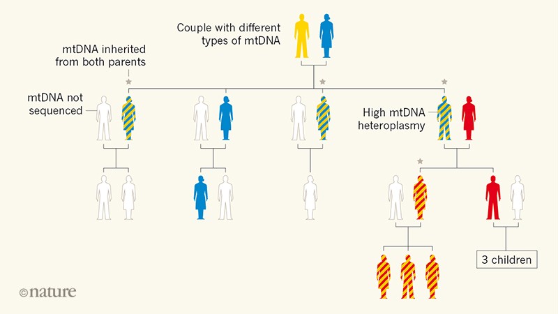
Figure 1 | Family tree revealing paternal inheritance of mitochondrial DNA (mtDNA). Luo et al.2
sequenced the mtDNA of several members of a family in which many
individuals had a high level of mtDNA heteroplasmy (the presence of
distinct genetic variants in the same cell). This mtDNA variability is
denoted by two colours in the same silhouette of an individual. The
analysis showed that some of the individuals with heteroplasmy had
inherited mtDNA from both of their parents, breaking the usual pattern
of exclusive maternal inheritance of mtDNA. Luo et al. suggest that the ability to inherit paternal mtDNA is a genetic trait.
To trace the origin of this mysterious mtDNA pattern, Luo et al.
extended their investigation to the previous generation. Sequencing of
the mtDNA of the boy’s maternal grandparents revealed an unexpected
contribution: his unusual mtDNA pattern seemed to be the product of
mtDNA from both grandparents. The authors went on to identify two
additional and unrelated families that had biparental mitochondrial
transmission. A similar scenario was previously observed in an
individual with mitochondrial disease who had a paternally inherited
mtDNA variant10. Together, these reports provide evidence for biparental mitochondrial inheritance in humans.
Human disease-causing mtDNA mutations were originally reported in 1988 (refs 6, 11)6,11, and more than 200 such mutations (see go.nature.com/2fucdqt) have been discovered since then, most of them occurring in a hetero-plasmic context7.
More-over, the estimated frequency of mutations of matrilineal mtDNA
has made it a useful and often-used tool in studies of ancestry and
evolution, as well as in forensic identification12.
Human mtDNA has also been a valuable tool in archaeology, because its
small size (16,569 base pairs) and circular form make it more resistant
to degradation than is nuclear DNA (which has around 3 billion base
pairs)13.
Given this long and multifaceted research history, why would paternal mtDNA have remained undetected? Luo et al.
suggest that mtDNA heteroplasmy is often overlooked in diagnostics when
it does not involve a disease-causing variant. Although this might be
true to some extent, it is a rather unsatisfactory explanation in this
era of deep DNA sequencing. Nevertheless, Luo and colleagues’ findings
should provoke a re-assessment of the extensive global mtDNA sequencing
data available, for those wishing to unearth further instances of
atypical heteroplasmy. If the paternal contribution to mtDNA is more
common than previously realized, this could alter some estimated timings
of human evolution, because these are often based on predictions of
mtDNA sequence variation under the assumption of exclusive maternal
inheritance.
Although biparental inheritance of mtDNA and
heteroplasmy coincided with disease symptoms in some of the individuals
studied by Luo et al., the authors’ data do not demonstrate a
causal link with disease. In fact, we cannot be certain that the study
participants have mitochondrial disease, because no specific
examinations to confirm this diagnosis are reported. Further study is
needed to identify more cases of potential paternal mtDNA inheritance,
and to determine the functional consequences of such heteroplasmy.
Notably, this knowledge is relevant to mitochondrial-donation therapy
(“three-parent babies”), which aims to prevent the transmission of
disease-causing mtDNA to offspring14, but which can also potentially generate individuals with two types of mtDNA, one from the donor and another from the mother.
Could
the amount of paternal mtDNA in a fertilized egg or developing embryo
be deliberately boosted to diminish the adverse effects of mutant
maternal mtDNA when this is present? This is an interesting option, but
still far from reality. In addition to evading elimination, paternal
mtDNA molecules would need to have a considerable replicative advantage
over maternal ones to reach meaningful proportions.
Will Luo and
colleagues’ findings affect the counselling of individuals carrying
disease-causing mtDNA mutations who are considering having children? Not
greatly, because paternal mitochondrial transmission seems to be
exceedingly rare in humans. At present, this discovery represents an
interesting conceptual breakthrough, rather than one that will directly
influence clinical practice.
Previous work15
has shown that mitophagy, the process by which cells ‘eat’ their own
mitochondria, has a role in the selective elimination of paternal
mitochondria. Given our rapidly expanding knowledge of mammalian
mitophagy in vivo16,
these rare instances of paternal mtDNA transmission might be attributed
to defective mitochondrial turnover.
The inheritance pattern of
paternal mtDNA in Luo and colleagues’ study suggests that a yet
unidentified gene on one of the autosomes (non-sex chromosomes) is
involved in eliminating paternal mitochondria. The families in whom
paternal mtDNA inheritance was observed provide an exciting opportunity
to decipher the signalling pathways that modulate paternal mitochondrial
elimination and prevent biparental mitochondrial transfer.
The
difference between eukaryotic and prokaryotic cells is that eukaryotic
cells are those which have a membrane-bound nucleus that contains
genetic material, as well as organelles that are also membrane-bound.
Whereas, prokaryotes are cells that don’t have a nucleus or
membrane-encased organelles.
An endothelial cell, an example of a eukaryotic cell. Photo: IP69.226.103.13 via Wikimedia Commons, CC 3.0All
living things are composed of cells. However, there are different kinds
of cells and cells are divided into one of two groups: prokaryotic
cells and eukaryotic cells. Eukaryotes (living things with eukaryotic
cell structures) and prokaryotes (living things with a prokaryotic cell
structure) are similar in many respects, but they have key differences
including different organelles that carry out different functions. As
previously mentioned, the major difference between prokaryotic and
eukaryotic cells is that prokaryotic cells lack a nucleus or organelles encased by a membrane, while eukaryotic cells have these features.
The distinction between eukaryotes and prokaryotes
is based upon two theories regarding the role of cells in biology. Cell
theory states that all living things are made out of cells, and
biogenesis theory proposes that all living cells came from other
existing cells, so cells cannot spontaneously arise from non-living
things (abiogenesis). For cells to comprise all the life we see on Earth
they must be able to organize proteins and other chemical compounds.
Individual cells are capable of keeping chemical processes isolated and
compartmentalized so one chemical process doesn’t interfere with another
chemical process, which runs the risk of disrupting the delicate
chemical balance of the cell and leading to cell death.
To keep
the chemical processes within a cell isolated and organized, cell
components stay enclosed within a membrane that serves as the barrier
between the interior of the cell and the cell’s environment. The
membrane of a cell is selectively permeable, meaning that the cell will
allow some chemical compounds into the cell and not others. The membrane
of the cell is made out of a phospholipid bilayer, two groups of lipids
arrayed in layers and facing opposite directions. Smaller compounds can
penetrate the phospholipid bilayer, but larger compounds must enter the
cell using a pore in the cell membrane.
The
cell membrane has various methods of regulating how chemicals move in
and out of the cell. Diffusion refers to the tendency of molecules at
high concentrations to distribute out to areas of lower concentration
until the concentrations across the two areas equalize. Osmosis is
similar to diffusion, but it refers to the movement of a solvent across a
boundary instead of a solute. This helps to equalize a solute that
can’t move through a boundary. Selective transport, the movement of
molecules within the cell, is handled by both membrane pumps and
membrane channels.
The Structure of Prokaryotes
Photo: Ali Zifan via Wikimedia Commons, CC 4.0Prokaryotes are made out of cells that don’t have a nucleus or organelles enclosed by membranes.
Since prokaryotic cells don’t have a nucleus, the DNA of the cell isn’t
locked within a nucleus. While prokaryotes don’t have a membrane-bound
nucleus the region that contains the DNA in a prokaryotic cell is a
central region referred to as the nucleoid. The DNA of prokaryotes is
overall less structured than the DNA found in eukaryotes. DNA structures
in prokaryotic cells are usually only a single loop, while the DNA
found in eukaryotic cells are bundled together in chromosomes.
A
eukaryotic cell typically has the following cell parts: cell wall,
plasma membrane, flagella, pili, cytoplasm, nucleoid, plasmid.
Another
key difference between prokaryote organisms and eukaryote organisms is
that most prokaryotes are single-celled organisms, though a few exist
that are made out of small collections of cells. Bacteria and archaea
are the two different forms of prokaryotes that scientists recognize.
Archaea are single-celled microorganisms that have genes and
chemical/metabolic pathways that are more similar to eukaryotes than
bacteria. Archaea lack a cell wall, whereas bacteria have a cell wall
composed of peptidoglycan. Many bacteria also have a polysaccharide
capsule. The function of the cell wall is to give the cell extra
protection from harmful compounds in the environment. The cell wall also
helps the cell maintains its shape and defends against dehydration. The
polysaccharide capsule lets the cell stick to surfaces in the
surrounding environment.
Prokaryotic cells often have tail-like
structures called flagella, which are used to propel the cell around.
The cells also have small tendrils called pili that are used to exchange
genetic material during a form of reproduction called conjugation.
The Structure of Eukaryotes
Eukaryotic organisms
are made out of cells that possess membrane-bound organelles and a
membrane-bound nucleus. The genetic material of eukaryotic organisms is
found within the nucleus of the cell and chromosomes are how the DNA is
organized within the nucleus. Eukaryotic cells comprise both
single-celled and multicellular forms of life. Eukaryotes compose the
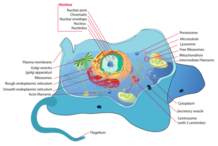
bodies of all protists, fungi, plants and animals. Photo:
By LadyofHats (Mariana Ruiz) – Own work using Adobe Illustrator. Image
renamed from Image: Animal cell structure.svg, Public Domain,
https://commons.wikimedia.org/w/index.php?curid=4266142Eukaryotic
cells display an impressive amount of diversity, and they are capable
of being many different shapes and sizes. The structure of a eukaryotic
cell is tied closely to its function. For example, the epithelial cells
that make up the human body are usually divided into three different
types: squamous, cuboidal, and columnar. Each of these different cell
types has a different form that enables it to carry out its respective
function.
Prokaryotic cells are almost always much smaller than eukaryotic cells.
The size of a prokaryotic cell is usually around 1 µm while the size of
animal cells and plants cells are usually between 10 to 100 µm. Because
Eukaryotic cells are much larger than prokaryotic cells they have
evolved special methods of transporting substances around the cell that
bacteria don’t have.
Summing Up:
Prokaryotic cells are
single-celled, have no membrane-bound nucleus, have a circular DNA
shape, reproduce asexually, are much smaller than eukaryotes, and are
divided into bacteria and archaea.
Eukaryotes can be multi-celled,
have a membrane-bound nucleus, have linear DNA, are capable of
reproducing sexually, are much larger than prokaryotes, and make up
plants, animals, protists, and fungi.