Carbonate concretions are known to contain well-preserved fossils and soft tissues. Recently, biomolecules (e.g.
cholesterol) and molecular fossils (biomarkers) were also discovered in
a 380 million-year-old concretion, revealing their importance in
exceptional preservation of biosignatures. Here, we used a range of
microanalytical techniques, biomarkers and compound specific isotope
analyses to report the presence of red and white blood cell-like
structures as well as platelet-like structures, collagen and cholesterol
in an ichthyosaur bone encapsulated in a carbonate concretion from the
Early Jurassic (~182.7 Ma).
The red blood cell-like structures are four
to five times smaller than those identified in modern organisms.
Transmission electron microscopy (TEM) analysis revealed that the red
blood cell-like structures are organic in composition. We propose that
the small size of the blood cell-like structures results from an
evolutionary adaptation to the prolonged low oxygen atmospheric levels
prevailing during the 70 Ma when ichthyosaurs thrived. The δ13C
of the ichthyosaur bone cholesterol indicates that it largely derives
from a higher level in the food chain and is consistent with a fish and
cephalopod diet. The combined findings above demonstrate that carbonate
concretions create isolated environments that promote exceptional
preservation of fragile tissues and biomolecules.
Introduction
Dinosaur
fossils, even with the most beautifully preserved anatomy, generally
lack soft tissues such as fibrous or cellular remains as well as
biomolecules or molecular fossils. However, over the last three decades,
several studies have shown that fragile tissues and molecules can be
preserved over surprisingly long periods of time (tens of millions of
years)1,2,3,4,5,6,7,8.
Heme-derived porphyrins were detected in a blood engorged mosquito from the Middle Eocene1.
More recently, red blood cell (RBC)-like structures, along with amino
acids associated with collagen-like fibres, were also found in 75
million-year-old dinosaur bones8.
The latter finding was remarkable considering the fact that the bone
fragments were not particularly well preserved, which is in agreement
with models suggesting that preservation of biomolecules and soft
tissues in the fossil record is more common than previously thought8,9,10,11. Collagen fibres were also reported in-situ in a 195 million-year-old dinosaur7.
Here, we investigated an ichthyosaur vertebra (Stenopterygius) of Lower Toarcian age (~182.7 Ma), which has been preserved through encapsulation in a carbonate concretion (Fig. 1).
The sample was collected from the renowned Posidonia Shale Konservat
Fossil Lagerstätte in SW-Germany. Ichthyosaurs thrived in the Mesozoic
era; they evolved following the largest mass extinction to have affected
life on our planet (during the Olenekian Stage of the Early Triassic,
between 251.1 Ma and 247.2 Ma)12,13 and became extinct at the end-Cenomanian (93.9 Ma)13.
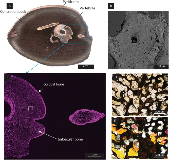
Figure 1
Morphology, mineralogy and chemical composition of ichthyosaur bones within a carbonate concretion. (A)
Photographic image of a polished section of the bone-containing
concretion. The vertebra served as the main nucleus which triggered the
microbial degradation processes leading to the concretion. The rim
contains a high amount of pyrite (observed by XRD, optical microscopy
and SEM) in contrast to the concretion body. (B) Backscattered electron image of a Haversian system, including Haversian canal, osteocytes and lamellae from the bone. (C)
Microbeam XRF elemental mapping of phosphorus (magenta) showing that
phosphorus is relatively more abundant in the bones than in the
concretion. The blue square represents the area where the Haversian
system was imaged. (D and E) Optical imaging on a thin section using ppl (D) and xpl (E) showing the minerals filling the porosity of the bone: equant sparry calcite (CaCO3) and barite (BaSO4). BaSO4
was identified by its mineral properties (clear colour and 90°
cleavages) and high birefringence as well as by elemental distribution
using microbeam XRF (Figure S1).
Generally,
during the Jurassic, ichthyosaur falls in shallow waters had low
preservation potential for tissues and biomolecules due to the presence
of a specialised consortium of degraders14. However, in the Lower Toarcian, when the Harpoceras falciferum
zone was deposited, the preservational environment in epicontinental
seas was excellent for tissues and biomolecules due to water column
stratification and strong euxinic conditions in the bottom waters15,16.
Under these euxinic conditions, organic matter-rich mudstones were
deposited and the diagenetic formation of carbonate concretions was
common17. Such carbonate concretions often contain fossils18,19 or, in some exceptional cases, even biomolecules20,21.
The
aim of this study was to investigate the potential of carbonate
concretions to preserve microscopic soft tissue and biomolecules from a
vertebra of the ichthyosaur Stenopterygius. A combined approach using in-situ
imaging techniques and molecular investigations was applied to study
this carbonate concretion and encapsulated fossil. Here, we report the
oldest RBC, white blood cell (WBC) and platelet-like structures,
>100 Myr older than in a previous report8 as well as the second oldest occurrences of collagen fibres7 and cholesterol20.
Results and Discussion
Encapsulation of an ichthyosaur vertebra in a concretion
A range of imaging techniques was applied to a polished section of the vertebra (Fig. 1A–E).
A selection of three-dimensional samples from the vertebra cortical and
trabecular bones was taken. Both cortical and trabecular bones display a
homogenous structure. Early mineralisation of concretions around the
decaying organic matter may occur within weeks or months22.
During this early encapsulation, the formation of a tight carbonate
cement prevented the bone from further microbial degradation and
inhibited exchange of fluids with the surrounding environment. The
concretion body is composed of microspar calcite and small (~10 µm)
dispersed euhedral crystals of pyrite. The outer rim of the concretion
is rich in pyrite. No septaria were observed within the concretion,
which further supports the limited post-depositional exchange with the
diagenetic environment. Therefore, early post mortem encapsulation led
to preservation of the bone tissue in the concretion.
Bone structure and elemental mapping
Microbeam
XRF mapping of phosphorus (P) showed that P is relatively more abundant
in the bone fragments than within the concretion (Fig. 1C), and helped to distinguish the cortical (i.e. compact) bone from the trabecular (i.e. spongy) bone. The high primary porosity (e.g. up to 65%) of vertebra bones has been reported previously in ichthyosaurs23. We calculated a porosity of the same range (estimated at ~60%) in the trabecular bone (Fig. 1C), where pores have been predominantly cemented by calcite (Fig. 1D and E). Elemental mapping (Ba, S) (Figure S1) and optical imaging (Fig. 1D and E) revealed a bone compartment cemented with trace element-enriched barite (BaSO4), a feature often observed in bones deposited under anoxic conditions where trace elements may be mobilised from a black shale24.
Examination
of the internal bone structure of the ichthyosaur, using backscattered
electron imaging, revealed remarkable preservation of fossilised 250
µm-diameter secondary osteons (Haversian system), known to be involved
in mature bone remodelling and renewal. Within the osteons, a number of
osteocytes and lamellae are visible (Fig. 1B).
Osteocytes play a predominant role in the synthesis of collagen and
regulate osteoblast function as well as biomineralisation of bones (e.g.25).
Red and white blood cells, platelets and collagen fibres in an ichthyosaur
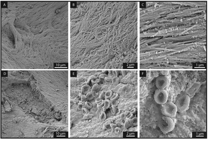
Scanning
electron microscopy (SEM) analyses were performed on samples from the
trabecular and cortical bones. Images were acquired after removal of the
carbonate filling the bone porosity, as described in Material and
Methods. SEM imaging of fossilised soft tissue in the trabecular bone
(Fig. 2) revealed intertwined elongated fibres (average width of 160 ± 1 nm; n = 88). These fibres show curved geometries and bundles (Fig. 2A–C) which, in size and orientation, resemble modern crocodile collagen (Figure S3).
These fibres also are within the diameter range (size comprised between
130 to 250 nm for 30 measurements) of collagen fibres reported in Late
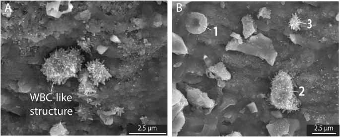
Cretaceous dinosaurs4,8. In close proximity to these collagen fibres, clusters of concave disks with an average size of 1.95 ± 0.21 µm (n = 75), closely resembling RBC-like structures reported from dinosaurs8, were observed (Fig. 2D–F). In addition to RBC-like structures, WBC- and platelet-like structures were identified (Fig. 3) based on morphological comparison with modern analogues26. However, all these blood cell-like structures are generally four to five times smaller than those identified in modern mammals27.
Figure 2
Secondary
electron images of the trabecular bone following the removal of sparry
calcite by light acetic acid treatment revealing exceptionally
well-preserved soft tissues. (A to C) Represent collagen fibres8 with increasing magnification. (D to F) Reveal RBC-like structures with increasing magnification.
Secondary
electron images of the trabecular bone following the removal of sparry
calcite by light acetic acid revealing soft tissues. (A) Presence of WBC-like structures. (B) 1) indicates a RBC-like structure, 2) indicates a WBC-like structure and 3) indicates a platelet-like structure.
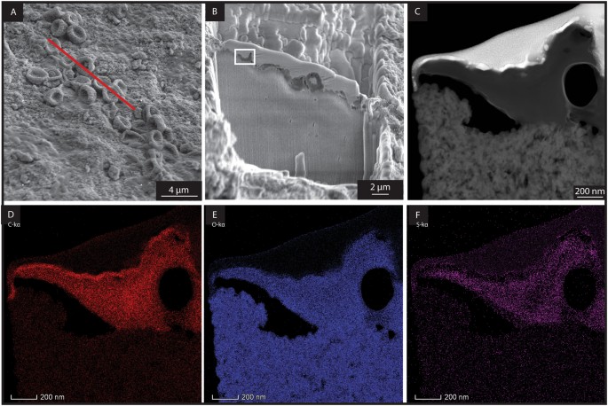
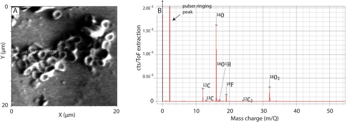
RBC-like structures were isolated and analysed by transmission electron microscopy (TEM), (Fig. 4)
which highlighted the presence of both carbon and oxygen in these
structures. Time of Flight Secondary Ion Mass Spectrometry (ToF-SIMS)
analyses of the RBC-like structures revealed the abundant light isotopes
of carbon (12C) and oxygen (16O), further supporting an organic origin (Figs 5 and S2).
Additional evidence for an organic origin is confirmed by the
identification of the polar compound Me,Et maleimide (3-ethyl,
4-methyl-pyrrole-2,5-dione) extracted from the bone. Indeed, Me,Et
maleimide is a known oxidative degradation product of heme and
chlorophyll pigments28. It is thus suggested that this maleimide likely derived from heme.
Figure 4
Area extracted by FIB-SEM for TEM analysis. (A)
Secondary electron image of the trabecular bone showing the presence of
RBC-like structures. The TEM foil was extracted from a cross-section
showed by the red line. (B) Secondary electron image taken during
TEM foil preparation showing the cross-section of the foil just prior
to lift-out. The white rectangle indicates the area selected for TEM
elemental mapping in (D,E and F). (C) TEM-HAADF image of a RCB-like structure. (D) Carbon (C) distribution of the RCB-like structure by TEM. (E) Oxygen (O) distribution of the RCB-like structure. (F) Sulfur (S) distribution in the RBC-like structure.
ToF -SIMS analysis of RBC-like structures in the ichthyosaur vertebra. (A)
Secondary image of the RBC-like structures by ToF-SIMS. The white
rectangle correspond to the area where the mass spectra was acquired. (B) Negative ions mass spectra showing the presence of C, O and Fluorine (F) specifically associated with the RBC-like structures.
Due
to their small size, the RBC-like structures could potentially be
interpreted as derived from bacteria. Here, we present several arguments
supporting a blood cell origin rather than a bacterial origin. All
RBC-, WBC-, and platelet-like structures were exclusively detected in
the vertebra bone. This is inconsistent with a bacterial origin, as
bacteria would be expected to be present in the vertebra as well as the
surrounding concretion (body and rim). In addition, all blood cell-like
structures were only revealed on the bones surfaces after removing the
carbonate filling the bone porosity. This suggests they were entombed
under the carbonate cement since it formed about 183 Ma ago, further
supporting that these blood cell-like structures cannot be the result of
recent bacterial colonisation. Furthermore, the RBC-like structures are
not simply deposited on the bone, but are locally fused into it (Fig. 2D–F), which is consistent with the fact that erythropoiesis (blood cell formation) occurs in medullar bones (e.g. vertebrae).
Lastly,
coccoid shaped bacteria are generally smaller (0.5–2 μm) than the
RBC-like structures observed here and they lack a concave shape. The
other major bacterial shapes (rods and vibrios) have absolutely no
resemblance with the shape of the RBC-like structures. For these
reasons, we conclude that the concave-shaped structures show
similarities with modern day RBCs. Similarly, the absence of hopanols
within the bone suggests that these structures are not of bacterial
origin. In addition, the dramatic variation in shape and size of RBCs
within a single class of modern animal (e.g. mammals) has been reported
since 1875 (as cited by29,30).
Since the extinction of the dinosaurs (~65 Ma), a rapid evolution and
diversification of mammalian species took place, colonising many
vacant ecological niches . This rapid evolution and diversification was
also reflected in the great variety of size and shape of RBCs in mammals29,30.
Similarly, during the Mesozoic era which lasted ~187 Myr, reptiles
reached their highest diversity and numerous species appeared and became
extinct. It seems highly possible that Jurassic reptiles could have
also presented diversity in their RBC shape as well as size, in order to
efficiently adapt to the surrounding paleoenvironmental conditions. We
therefore propose that the small size of these blood cell-like
structures observed therein is related to evolutionary adaptation to
environmental conditions.
Evolutionary adaptation to environmental conditions
Ichthyosaurs
evolved during an episode typified by low atmospheric oxygen levels,
lasting over 70 million years from the Early Triassic to the Lower
Jurassic31. We suggest that under the prolonged low oxygen levels in the atmosphere32,33,34, small RBCs could have been favoured because the surface to volume ratio35
provides a more efficient oxygen transport and diffusion. For example,
mammals living at high altitude have been shown to have excellent
adaptation to low oxygen levels based on abundant RBCs of small size35. The “bowl-like” shape of the cells resembling RBCs (i.e. stomatocytes) has been widely reported in disease-related studies of mammalian species with anucleated RBCs36,37. However, the study of blood in reptiles is limited, which makes the interpretation of reptilian hematologic data challenging38,39.
We
hypothesise that the fossil occurrence of small RBC-like structures in
ichthyosaurs could be consistent with an oxygen-depleted
palaeoenvironment and evolutionary adaptation. This adaptation is
supported by the occurrence of RBC-like structures of similar size in
terrestrial dinosaurs8. Although oxygen concentrations reached today’s levels during the Late Cretaceous40,
most of dinosaurs’ evolution took place during prolonged periods of low
oxygen levels and they lived under the same atmospheric conditions as
the ichthyosaurs. In modern fish, RBCs size has been shown to be
inversely proportional to aerobic swimming ability41. Moreover, a correlation between small RBCs size and high rate of metabolism has also been demonstrated in modern geckos42,43. With respect to adaption, we emphasize that Stenopterygius is considered to have been one of the fastest marine predators of its time44, its cruising speed equivalent to that of modern day dolphin and with a similar morphology45. A high degree of RBC aggregation has previously been reported in modern higher athletic species46. This metabolic adaptation could potentially explain the clustering of the small RBC-like structures observed in this Stenopterygius.
In order to sustain the metabolism required for high-speed pursuit
predators, the muscular tissue must have been highly efficient and have
been supported by a complex blood circulation system, adapted to
low-oxygen environment, to provide sufficient oxygen to the lungs of the
ichthyosaurs. Given that the bone studied is a medullary bone (i.e.
vertebra), it would yield sufficient bone marrow (see below) to
synthesise RBCs. Based on their small size, the fossilised RBC-like
structures indicate a fast and efficient oxygen diffusion into the
cells, allowing for high pursuit speed and thus providing competitive
advantage over slower moving prey.
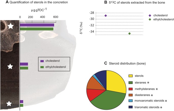
Cholesterol in an ichthyosaur
Besides
fossilised RBC-, WBC- and platelet-like structures, the ichthyosaur
bone contained elevated concentrations of the biomolecule cholesterol
(565 µg/g TOC, Fig. 6 and Table S1). It was previously reported that free cholesterol is relatively abundant in the bone marrow47
supporting the high amount of neutrally extracted free bone cholesterol
in our sample. The bone cholesterol differed in its isotopic carbon
composition (−28.9‰ VPDB) compared to ethylcholesterol (−34.6‰ VPDB;
Fig. 6). The isotopic discrepancy between these two sterols supports different origin(s). The 13C
enrichment of the cholesterol by 5.7‰ VPDB indicates that it largely
derives from a higher level in the food chain and corroborates a fish
and cephalopod diet of the ichthyosaur48,49. The 13C
isotopic composition of the ethylcholesterol is consistent with a
source from phytoplankton in the ancient water column. Recently, soft
tissue of a crustacean inside a Devonian concretion from the Gogo
Formation (Canning Basin, Western Australia) was reported to contain an
entire diagenetic continuum of organic molecules with the remarkable
co-occurrence of biomolecules and geomolecules, from sterols to
triaromatic steroids (including sterenes and diasterenes)20.
The exceptional preservation of these compounds was attributed to rapid
encapsulation by microbially-mediated and eogenetic processes. In our
study, steroid end-products of diagenesis were also identified in
association with the vertebra (Fig. 6C).
However, the absence of sterenes and diasterenes suggests the formation
of the concretion within the sediments (corroborated by the
preservation of slightly disturbed sedimentary bedding) and was not
initiated in the water column20,21. The Posidonia Shale Formation and the Gogo Formation concretions were both formed under similar euxinic (H2S-rich) conditions and are well known Fossil–Lagerstätten.
Figure 6
Steroid distribution and compound specific δ13C values. (A)
Sterol distribution within the bone and concretion, showing high
concentrations of cholesterol (565 µg/g TOC) and ethylcholesterol
(523 µg/g TOC) and lower concentrations in the concretion body and rim. (B) δ13C
values (‰ VPDB) of sterols associated with the bone (cholesterol:
−28.9 ± 0.4‰ VPDB; ethylcholesterol: −34.6 ± 0.4‰ VPDB) from the
concretion. The error bars are contained within the symbol. (C)
Relative proportion of compound classes within the fossil, dominated by
sterols and steranes representing the end-members of the diagenetic
sequence. *Diagenetic end products.
Tight
encapsulation of a Jurassic ichthyosaur bone in a diagenetically formed
carbonate concretion allowed for the fossilisation of structures
showing resemblance to modern day bone collagen, RBCs, WBCs and
platelets with a micron-scale preservation. Microanalysis revealed that
the RBC-like structures are enriched in organic material.
The
rapid encapsulation of the vertebra also led to the preservation of
cholesterol, largely derived from the vertebra bone, over a period of
~182.7 million years. These observations highlight the development of a
closed environment within carbonate concretions. In such cases,
carbonate concretions preserve fossils (structural and cellular) and
biomolecules, as well as molecular fossils with an excellent level of
detail. This well-preserved primary biomaterial suggests that carbonate
concretions could play a major role in the investigation of the
palaeobiology of extinct species and in understanding the evolution of
life. Based on the small size of the RBCs and their associated high
oxygen diffusion capacity, we hypothesise that ichthyosaur must have
followed a high-energy life style as a pursuit predator. This would
distinguish its hunting strategy from that of an ambush predator life
style as assumed for plesiosaurs.
Material and Methods
Geological settings and sampling
The
investigated sample was recovered from the Toarcian Posidonia Shale
Formation at the HOLCIM Cement quarry of Dotternhausen (SW-Germany). The
concretion was collected shortly after blasting within the quarry and
stored in the dark at room temperature before a transverse slice was cut
across the oval shaped concretion (Fig. 1A), just before analyses.
Following
the global Toarcian transgression, the black shale host sediment was
deposited in the SW-German sub-basin, on the epicontinental Western
Tethyan Shelf50,51,52.
The sub-basin experienced high algal productivity coupled with
restrictions in water circulation leading to stratification of the water
column and development of anoxic to euxinic bottom waters, which
favoured the deposition of organic matter-rich black shales16,51,52.
Sample preparation
Three
1 cm-slices were cut from the central area of the lens shaped
concretion, perpendicular to the horizontal bedding. One slice was
polished for microbeam X-Ray Fluorescence (XRF) (Bruker M4 TORNADOTM)
elemental mapping. A thin section of the vertebra was prepared from the
second slice. From the third slice, three samples were taken from i)
vertebra, ii) concretion body presenting sedimentary bedding and iii)
concretion rim for organic geochemical and compound specific isotope
analyses (CSIA). Each sample was cleaned in an ultrasonic bath using a
mixture of dichloromethane: methanol (DCM: MeOH) at 9:1 (v/v) (three
times × 20 min) to remove any surface contaminants. Fossil bone samples
were crushed and mm-sized pieces of bone were collected and treated with
1 M acetic acid solution immediately prior to SEM imaging. The
remainder of the samples was pulverised using a Rocklabs benchtop ring
mill (BTRM) in a pre-annealed zircon mill. Pre-annealed quartz sand was
pulverised and analysed as a procedural blank for organic geochemical
techniques. Mineralogy was determined using aliquots of pulverised
samples for X-ray diffraction (XRD).
A modern crocodile leg sample
was obtained from Mahogany Creek Distributors (Perth, Australia). The
bones were cut and isolated from the flesh. A sample was then left to
dry in the oven for 24 h (50 °C, below the denaturation threshold of
native hydrated collagen fibrils53) and treated with concentrated H2O2 (48 h). The oxidised flesh and bone marrow were removed using forceps and the bone was left to dry at room temperature.
Mineralogy
XRD
analyses on powdered samples were performed using a Bruker-AXS D8
Advance Diffractometer with CuKα radiation and a LynxEye position
sensitive detector. The data were collected from 7.5 to 90° 2Ө, with a
nominal step size of 0.015° and a collection time of 0.7 seconds per
step. Crystalline phases were identified using the Search/Match
algorithm, DIFFRAC.EVA 3.1 (Bruker-AXS) to search the Powder Diffraction
File.
Imaging methods
Porosity estimation
The
estimation of the trabecular porosity of the ichthyosaur vertebra has
been determined through digital point counting on a recursive grid to
two times 200 points and stabilisation of the point count distribution
plot. The overall trabecular porosity evaluated was estimated at 59.5%.
Microbeam XRF mapping
A
Bruker M4 TORNADO™ Micro-XRF equipped with a rhodium target X-ray tube
operating at 50 kV and 500 nA and an XFlash® silicon drift X-ray
detector was used for elemental mapping of the polished slice of the
Toarcian concretion samples. Maps were created using a 25 µm spot size
on a 25 µm raster with dwell time of 5 ms per pixel.
Scanning Electron Microscopy (SEM)
Both
modern crocodile and fossil ichthyosaur bone samples were coated using a
Quorum Q150T ES coating unit. A carbon layer of approximately 25 nm was
applied and as samples were charging, an additional thin coating of
gold (3–5 nm) was applied.
The bone samples were examined using a
Tescan Mira-3 Field Emission Gun Scanning Electron Microscope (FEG-SEM).
The instrument was operated with an accelerating voltage 5 kV and a
beam current of approximately 200 pA. The images acquired were collected
using an Everhart-Thornley Secondary Electron (SE) detector.
Focused Ion Beam Scanning Electron Microscope (FIB-SEM)
The
sample was examined using a Tescan Lyra FIB-SEM. A small fragment of
the bone was mounted onto an aluminium stub and coated with gold. A
cross-sectional lamella covering a number of RBC-like structures was
extracted using standard FIB-SEM lift out techniques, mounted onto a
copper grid and thinned to ~100 nm, followed by a low kV (2 kV) ‘clean
up’ routine to remove surface damage.
Time of flight secondary ion mass spectrometry (ToF-SIMS)
ToF-SIMS
was performed during microstructural analysis using a Tescan Lyra. The
instrument is fitted with a TOFWERK ToF-SIMS detector and uses the Ga+
ion beam from the FIB as the primary ion source. Analysis was performed
over a 20 µm × 20 µm area to a depth of approximately 400 nm using a
20 kV primary ion energy and a current of 500 pA. Negative ions were
collected and a mass spectrum was derived from a volume containing only
RBC-like structures to reveal their chemical composition.
Transmission Electron Microscopy (TEM)
Microstructural
analysis and elemental mapping of the FIB-SEM prepared lamella was
carried out using high angle annular dark field scanning transmission
electron microscopy (HAADF-STEM, FEI Talos F200x TEM/STEM with Super-X
EDS Detectors) at 200 kV.
Lipid biomarker analyses
Toarcian
samples were subject to Soxhlet extraction with a mixture of DCM:MeOH
(9:1, 72 hrs). Activated copper turnings were added to remove elemental
sulfur. An aliquot of each total lipid extract was adsorbed onto
activated silica gel (160 °C, >24 hrs). Each aliquot was then
separated using column chromatography with a small column containing
activated silica gel (5 cm × 0.5 cm i.d.) into five fractions. i) The
aliphatic hydrocarbon fraction was eluted using 2 mL of n-hexane; ii) the aromatic hydrocarbon fraction was eluted with 2 mL n-hexane:DCM
(4:1); iii) the ketone and fatty acid methyl esters fraction was eluted
with 2 mL DCM; iv) the sterol fraction was eluted with 2 mL DCM:ethyl
acetate (4:1) and v) the polar fraction was eluted using DCM:MeOH (7:3).
Sterols
were derivatised using bis(trimethylsilyl)-trifluoroacetamide (BSTFA)
and anhydrous pyridine (for 100 µg, 60 µL BSTFA, 40 µL pyridine) heated
at 70 °C for 30 min and dried under a nitrogen purge. The fractions were
dissolved in n-hexane and analysed using gas chromatography-mass
spectrometry (GC-MS). Semi-quantitative analyses of the sterol
fractions were carried out using external calibration with a derivatised
cholesterol standard. Procedural blanks were performed throughout.
GC-MS
analyses were performed using a Hewlett Packard 6890 gas chromatograph
(GC) interfaced with a Hewlett Packard 5973 mass selective detector. The
GC was equipped with a split/splitless injector and a DB-5 capillary
column (60 m × 0.25 mm i.d. coated with a 0.25 µm film thickness). The
initial oven temperature (50 °C) was increased at a rate of 6 °C/min
until reaching the final temperature (320 °C), initial and final hold
times were 1 minute and 24 minutes, respectively. Ultra-high purity
helium was used as a carrier gas at a constant flow (1.1 mL/min). The MS
detector was operated at 70 eV (full scan) from 35–650 Da.
The detailed procedure used for maleimide purification is reported elsewhere54.
In brief, polar fractions were purified by thin layer chromatography
(TLC) using DCM:ethyl acetate (EtOAc) (4:1, v-v), along with a reference
compound H,H maleimide (Sigma Aldrich) used as a retention standard.
The band between retention factor (Rf) 0.6 and 0.9 (containing the
maleimides) was recovered by elution with EtOAc over a small silica gel
column.
Derivatisation with N-(tert-butyldimethylsilyl)-N-methyl
trifluoroacetamide (MTBSTFA) in pyridine was performed to obtain
tert-butyldimethylsilyl (TBDMS) derivatives of maleimides (e.g.28,54). TBDMS derivatives of maleimide in n-hexane
were analysed by GC-MS using an Agilent HP 6890 GC system equipped with
an Agilent DB-5MS column [60 m × 0.25 mm i.d. × 0.25 μm f.t.] coupled
to an Agilent 5973 Mass Selective Detector operated at 70 eV. The
temperature program for both instruments was 40 °C (1 min isothermal),
40 °C to 100 °C at 10 °C/min, 100 °C to 320 °C at 4 °C/min, isothermal
at 320 °C for 30 min. Helium was used as carrier gas (1.2 mL/min). The
maleimide was identified based on their mass spectrum, retention times
and elution order by comparison with other published work (e.g.29,53).
CSIA
was performed on a Thermo Finnigan Delta V mass spectrometer coupled to
an Isolink GC. A pure cholesterol standard (underivatised and
derivatised) was analysed in order to calculate the δ13C of the additional methyl-groups from BSTFA54. Samples were run as triplicates and the δ13C values of the parent compounds were corrected for the isotopic composition from the methyl-groups of the BSTFA54. A CO2 reference gas standard with a known δ13C value was introduced during CSIA to determine the δ13C values of the sterols. The δ13C
are reported in per mil (‰) relative to the international Vienna Peedee
Belemnite standard (VPDB); the values reported have a standard
deviation below 0.4‰VPDB for at least 3 analyses.
Availability of materials and data
The
datasets generated during and/or analysed during the current study are
available from the corresponding author on reasonable request.
Additional information
Publisher's note: Springer Nature remains neutral with regard to jurisdictional claims in published maps and institutional affiliations.
References
1.
Greenwalt,
D. E., Goreva, Y. S., Siljeström, S. M., Rose, T. & Harbach, R. E.
Hemoglobin-derived porphyrins preserved in a Middle Eocene
blood-engorged mosquito. Proc. Natl. Acad. Sci. USA110, 18496–18500 (2013).
Schweitzer,
M. H., Wittmeyer, J., Horner, J. R. & Toporski, J. K. Soft-tissue
vessels and cellular preservation in Tyrannosaurus rex. Science307, 1952–1955 (2005).
Schweitzer,
M. H., Wittmeyer, J. L. & Horner, J. R. Soft tissue and cellular
preservation in vertebrate skeletal elements from the Cretaceous to the
present. Proc. Biol. Sci.274, 183–197 (2007).
Schweitzer,
M. H., Johnson, C., Zocco, T. G., Horner, J. R. & Starkey, J. R.
Preservation of biomolecules in cancellous bone of Tyrannosaurus rex. J. Vertebr. Paleontol.17, 349–359 (1997).
Asara,
J. M., Schweitzer, M. H., Freimark, L. M., Phillips, M. & Cantley,
L. C. Protein sequences from mastodon and Tyrannosaurus rex revealed by
mass spectrometry. Science316, 280–285 (2007).
Lee, Y.-C. et al. Evidence of preserved collagen in an Early Jurassic sauropodomorph dinosaur revealed by synchrotron FTIR microspectroscopy. Nat. Commun.8, 14220 (2017).
Schweitzer, M. H. et al. A role for iron and oxygen chemistry in preserving soft tissues, cells and molecules from deep time. Proc. Biol. Sci.281, 20132741 (2014).
Motani, R. Evolution of Fish-Shaped Reptiles (Reptilia: Ichthyopterygia) in Their Physical Environments and Constraints. Annu. Rev. Earth Planet. Sci.33, 395–420 (2005).
Fischer,
V., Bardet, N., Benson, R. B. J., Arkhangelsky, M. S. & Friedman,
M. Extinction of fish-shaped marine reptiles associated with reduced
evolutionary rates and global environmental volatility. Nat. Commun.7, 1–11 (2016).
Berner,
Z. A., Puchelt, H., Nöltner, T. & Kramar, U. Pyrite geochemistry in
the Toarcian Posidonia Shale of south-west Germany: Evidence for
contrasting trace-element patterns of diagenetic and syngenetic pyrites.
Sedimentology60, 548–573 (2013).
Schwark,
L. & Frimmel, A. Chemostratigraphy of the Posidonia Black Shale,
SW-Germany II. Assessment of extent and persistence of photic-zone
anoxia using aryl isoprenoid distibutions. Chem. Geol.206, 231–248 (2004).
Melendez, I. et al. Biomarkers reveal the role of photic zone euxinia in exceptional fossil preservation: An organic geochemical perspective. Geology41, 123–126 (2013).
Yoshida, H. et al. Early post-mortem formation of carbonate concretions around tusk-shells over week-month timescales. Sci. Rep. 1–7 https://doi.org/10.1038/srep14123 (2015).
23.
Lopuchowycz, V. B. & Massare, J. A. Bone Microstructure of a Cretaceous Ichtyosaur. Paludicola3, 139–147 (2002).
Leduc, T. Diagenesis of the fossil bones of Iguanodon bernissartensis from the Iguanodon sinkhole. In Bernissart dinosaurs and Early Cretaceous terrestrial ecosystems (ed. Godefroit, P.) 111–136 (Indiana University Press, 2012).
25.
Sanchez,
S., Tafforeau, P. & Ahlberg, P. E. The humerus of Eusthenopteron: a
puzzling organization presaging the establishment of tetrapod limb bone
marrow. Proc. Biol. Sci.281, 20140299 (2014).
Bergman, R. A., Afifi, A. K., Heidger, P. M. & D’Alessandro, M. P. Atlas of Microscopic Anatomy: A functional Approach. http://www.anatomyatlases.org/.
27.
Gregory, T. R. The Bigger the C-Value, the Larger the Cell: Genome Size and Red Blood Cell Size in Vertebrates. Blood Cells, Mol. Dis.27, 830–843 (2001).
Grice, K. et al. Maleimides (1H-pyrrole-2,5-diones) as Molecular Indicators of Anoxygenic Photosynthesis in Ancient Water Columns. Geochim. Cosmochim. Acta60, 3913–3924 (1996).
Yamaguchi,
K., Jürgens, K. D., Bartels, H. & Piiper, J. Oxygen transfer
properties and dimensions of red blood cells in high-altitude camelids,
dromedary camel and goat. J. Comp. Physiol. B157, 1–9 (1987).
Reagan,
W. J., Irizarry Rovira, A. R. & DeNicola, D. B. Veterinary
hematology. Atlas of common domestic and non domestic species.
(Wiley-Blackwell, 2008).
37.
Stewart, G. W. The hereditary stomatocytosis and allied conditions’: inherited disorders Na+ and K+ transport. In Red Cell Membrane Transport in Health and Disease (eds Ingolf, B. & Clive, E. J.) 511–523 (Springer Berlin Heidelberg, 2003).
38.
Nardini, G., Leopardi, S. & Bielli, M. Clinical hematology in reptilian species. Vet. Clin. North Am. Exot. Anim. Pract.16, 1–30 (2013).
Claver,
J. A. & Quaglia, A. I. E. Comparative Morphology, Development, and
Function of Blood Cells in Nonmammalian Vertebrates. J. Exot. Pet Med.18, 87–97 (2009).
Tappert, R. et al. Stable carbon isotopes of C3 plant resins and ambers record changes in atmospheric oxygen since the Triassic. Geochim. Cosmochim. Acta121, 240–262 (2013).
Starostová,
Z., Kubička, L., Konarzewski, M., Kozłowski, J. & Kratochvíl, L.
Cell Size but Not Genome Size Affects Scaling of Metabolic Rate in
Eyelid Geckos. Am. Nat.174, E100–E105 (2009).
Starostová,
Z., Konarzewski, M., Kozłowski, J. & Kratochvíl, L. Ontogeny of
Metabolic Rate and Red Blood Cell Size in Eyelid Geckos: Species Follow
Different Paths. PLoS One8, 21–23 (2013).
Popel,
A. S., Johnson, P. C., Kameneva, M. V. & Wild, M. A. Capacity for
red blood cell aggregation is higher in athletic mammalian species than
in sedentary species. J. Appl. Physiol.77, 1790–4 (1994).
Fröbisch,
N. B., Fröbisch, J., Sander, P. M., Schmitz, L. & Rieppel, O.
Macropredatory ichthyosaur from the Middle Triassic and the origin of
modern trophic networks. Proc. Natl. Acad. Sci. USA110, 1393–7 (2013).
Druckenmiller,
P. S., Kelley, N., Whalen, M. T., McRoberts, C. & Carter, J. G. An
Upper Triassic (Norian) ichthyosaur (Reptilia, Ichthyopterygia) from
northern Alaska and dietary insight based on gut contents. J. Vertebr. Paleontol.34, 1460–1465 (2014).
Ziegler,
P. A. EUROPE: Permian to Recent Evolution: Jurassic. in Encyclopedia of
geology (eds Selley, R. C., Cocks, L. R. M. & Plimer, I. R.)
106–112 (Elsevier, 2005).
51.
Röhl,
H., Schmid-Röhl, A., Oschmann, W., Frimmel, A. & Schwark, L. The
Posidonia Shale (Lower Toarcian) of SW-Germany: an oxygen-depleted
ecosystem controlled by sea level and palaeoclimate. Palaeogeogr. Palaeoclimatol. Palaeoecol.165, 27–52 (2001).
Schmid-Röhl,
A., Röhl, H., Oschmann, W. & Frimmel, A. Palaeoenvironmental
reconstruction of Lower Toarcian epicontinental black shales (Posidonia
Shale, SW Germany): global versus regional control. Geobios35, 13–20 (2002).
Jones,
D. M., Carter, J. F., Eglington, G., Jumeau, E. J. & Fenwick, C. S.
Determination of δ13C values of sedimentary straight chain and cyclic
alcohols by gas chromatography/isotope ratio mass spectrometry. J. mass Spectrom.20, 641–646 (1991).
The
authors thank Geoff Chidlow, Alex Holman and Marieke Sieverding for
their technical support with GC-MS and GC-irMS analyses. Sebastian’s
Butchers (Kalamunda, WA) is thanked for cutting and de-fleshing the
crocodile bones. Grice and Plet thank the Australian Research Council
(ARC) for an ARC-DORA grant (awarded to Grice: DP130100577) and for
Plet’s PhD stipend. Plet acknowledges Curtin University for a Curtin
International Postgraduate Research Training Scholarship and The
Institute of Geoscience Research (TIGeR) for a top-up scholarship, as
well as the European Association of Organic Geochemist (EAOG) for a
travel award to the organic geochemistry workgroup at Christian Albrecht
University (Germany). The microanalysis work was supported by the
Science and Industry Endowment Fund. Dr Zakaria Quadir is thanked for
his assistance with the TEM analysis. Schwark acknowledges support by
DFG-grant (Schw554/23-1,2) as well as permission to conduct and
assistance with field work by M. Jaeger, who identified the Stenopterygius sp. and A. Schmidt-Roehl, HOLCIM, Dotternhausen. The species was identified by comparison with other curated specimens of Stenopterygius.
Author information
Affiliations
WA-Organic
and Isotope Geochemistry, Department of Chemistry, The Institute for
Geoscience Research, Curtin University, Curtin, WA, 6845, Australia
Chloé Plet
, Kliti Grice
, Anais Pagès
, Marco J. L. Coolen
& Lorenz Schwark
CSIRO CESRE, Mineral resources, Kensington, WA, 6151, Australia
Anais Pagès
& Michael Verrall
Department of Organic Geochemistry, Institute of Geoscience, Christian Albrechts University, Kiel, 24118, Germany
Wolfgang Ruebsam
& Lorenz Schwark
Advanced Resource Characterisation Facility, John de Laeter Centre, Curtin University, Curtin, WA, 6845, Australia
William D. A. Rickard
Contributions
C.P.:
acquisition of data, analysis of data, interpretation and major writing
of manuscript. K.G.: conception and design, acquisition of data,
analysis of data, interpretation and major writing of manuscript and ARC
grant funding. A.P.: acquisition of data, and minor edits to the
writing of the manuscript. M.V.: acquisition of data, and minor edits to
the writing of the manuscript. M.J.L.C.: Interpretation and major
writing of manuscript. W.R.: acquisition of study material and data and
minor edits to the writing of the manuscript. W.R.: acquisition of data,
and minor edits to the writing of the manuscript. L.S.: conception and
design, acquisition of study material and data, analysis of data,
interpretation and major writing of manuscript.
Competing Interests
The authors declare that they have no competing interests.
Nenhum comentário:
Postar um comentário
Observação: somente um membro deste blog pode postar um comentário.