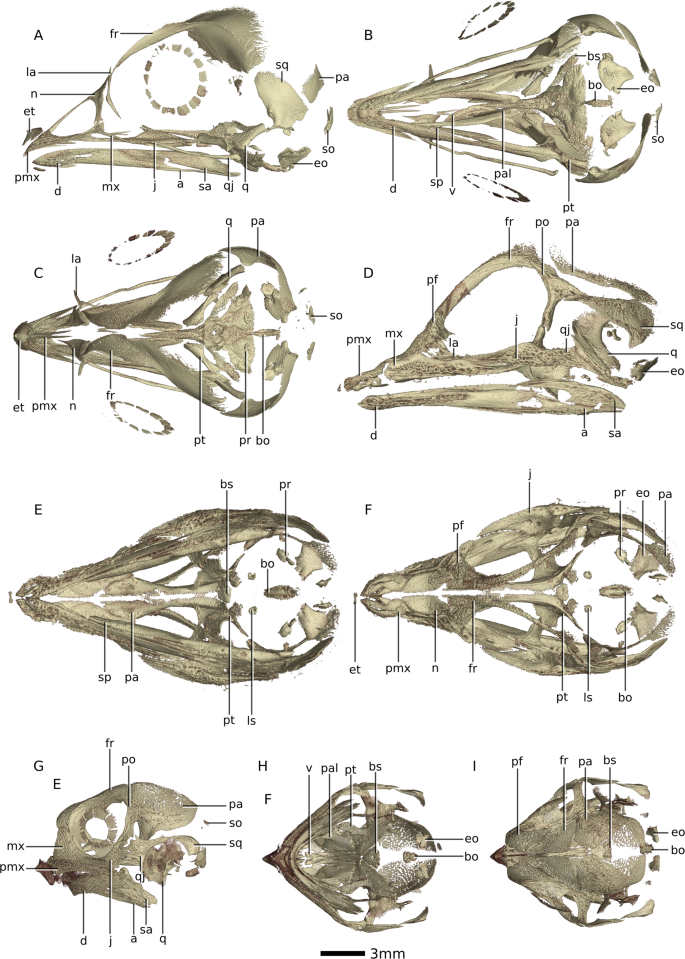
Our
results show that both the sequence and to a certain extent, the
relative timing of cranial ossification are conserved between
Gallus gallus,
Crocodylus niloticus,
Centrochelys sulcata and
Pogona vitticeps. This sequence agrees with previous work using different methods done on larger phylogenetic samples (including mammals)
31,32,54. In general, this order follows the aforementioned modules described in the event-pair cracking study
32
(i.e. jaw bones, the palatal bones, the bones forming the orbit, the
skull roof bones and the braincase bones), however some bones ossify
later than the rest of their respective modules (such as the coronoid,
the laterosphenoid, the articular and the palpebral).
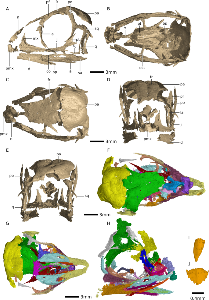
Based on the
level of ossification of the basisphenoid, frontals, parietals, palate,
and quadrate, as well as the absence of the remaining braincase bones
and the articular, we hypothesize that the
Massospondylus carinatus embryos are approximately 60% through their incubation period (56% or 48 days out of 85 for crocodiles
55;
67% or 12 days out of 21 for chickens; 61% or 61 days out of 100 for
spurred tortoises and 74% or 48 days out of 65 for bearded dragons
56) (see Fig.
2).
In our analysis, the bearded dragon was not scored between 36 days and
48 days in the incubation period (or 55–74%), as this gap was not
illustrated in the literature. This comparison therefore has lower
precision than the others and is considered with caution. Although at
this relative age we would expect the
Massospondylus carinatus
embryos to have an onset of ossification in the rest of the braincase
bones (i.e notably exoccipital and basioccipital but also possibly the
supraoccipital, prootic and laterosphenoid), the extant taxa present a
small ossification centre for these bones at this developmental
percentage. It is therefore possible that these had not started to
ossify yet in the dinosaur embryos, that the small ossification centres
did not preserve, or that they were below the resolution threshold in
our phase -contrast -based SRµCT data. This indicates that the embryos
are certainly not much more developed than 60% through their incubation
period, as these braincase bones would otherwise be more ossified and
visible. In
Gallus gallus,
Crocodylus niloticus, Centrochelys sulcata, and
Pogona vitticeps, these braincase bones are all at stage-code 2 at
approximately 70–75% through the incubation period (day 15, day 55, day
70 and day 60 respectively, see Table
S3). This puts an upper limit on the developmental percentage of the
Massospondylus carinatus embryos because stage-code 2 ossification is readily seen in our scan in other bones of the skull.
Our hypothesized relative developmental percentage of the
Massospondylus carinatus embryos indicates that they are earlier in development than previously thought
46.
This makes them some of the ontogenetically youngest dinosaur embryos
known. All other dinosaur embryos in the literature with ontogenetic age
estimates are hypothesized as being in the last third of their
development or near hatching
35,36,37,38,39.
However, analysing the latter using micro computed tomography scans and
our stage-code method could reveal that some of them are younger than
the
Massospondylus carinatus embryos presented here.
Our hypothesis for the developmental percentage of the
Massospondylus carinatus embryos is corroborated by the presence of both null-generation teeth and crowns with adult tooth morphology (see Fig.
1). Although many dinosaur embryos have been found to have teeth
36,37,38,40,
in all cases the reported morphology is most similar to adult teeth. To
our knowledge, null-generation teeth have not been reported in
dinosaurs, nor have null-generation and adult teeth been reported in a
single embryo.
Troodon teeth have cylindrical roots,
linguobuccaly compressed and mesiodistally elongated crowns. These were
hypothesized as being early developmental casts of unossified teeth
35.
Maiasaura
embryos preserve different generations of teeth, however the budding
teeth are hypothesized to grow in the form of the larger teeth
preserved. These are therefore replacement teeth and are similar in
morphology
57.
Null-generation teeth form during embryonic development in several reptile species
58,59,60.
These non-functional teeth are small, unicuspid (even if adult tooth
morphology is multicuspid), and possess little or no enamel. They are
either resorbed into the jaw or shed into the oral cavity
59.
Little information is available on null-generation teeth in living
saurian taxa. However, in geckos, the null-generation teeth appear at
about 23% through the incubation period. By 66% into the incubation
period, null-generation teeth are present in half of the tooth
positions, while the other half are formed by the first functional teeth
(null-generation teeth are therefore not a prerequisite for the
formation of adult tooth morphologies). By the end of the incubation
period, the first set of functional teeth have started to be resorbed
while the second set of functional teeth start moving orally
58. All null-generation teeth are replaced by functional teeth during embryonic development
61. The simple conical tooth morphology seen in the
Massospondylus carinatus embryos probably represent null-generation teeth for
Massospondylus carinatus,
which will get resorbed or shed into the oral cavity before hatching.
The abundance of these null-generation teeth (approximately half of the
teeth preserved, see Fig.
S14) correlate well with the 60% in incubation period estimated by the bone ossification.
Our
stage-code method provides a simple, relatively precise, repeatable
estimate of the developmental percentage in other extinct and extant
saurian embryos. Such estimates are of broad utility when including
embryos in broader ontogenetic studies. For example, the early
developmental percentage of the
Massospondylus carinatus embryos
suggests caution against uncritical use of limb measurements as part of
allometric studies, e.g., postural determination. Coupling these
developmental percentage estimates with incubation period
7
also provides better insight into the life histories of dinosaurs and
could potentially enable more precise future studies of how birds, for
example, decreased their incubation periods.
Although Reisz
et al.
(2010) identified embyronic skeletal material in five of the eggs that
comprise BP/1/5347a, our results show that only three of the seven eggs
contain embryonic material. Several factors have been hypothesized to
have an effect on clutch viability in extant taxa including
environmental conditions, eggshell structure, predation and microbial
contamination
62,63. However the patterns of embryonic mortality are poorly understood
63.
There are many potential reasons for fossil eggs being empty,
including: a high number of infertile eggs; high levels of early
mortality before ossification of the skeleton; leakage from broken eggs
during fossilisation; or that the clutch represents two or more clutches
laid at different times (Deeming, pers. com.). Given our sample size,
we cannot assess any of these critically at this time. However, the
three preserved embryos do not differ substantially in their
ossification, indicating that they are therefore at similar
developmental percentages and are probably from the same clutch. The
first and second embryos show some slight differences in level of
ossification. The Embryo 1 has a partial basipterygoid process that has
started to ossify as well as a quadrate. Embryo 2 does not have either
of these elements ossified. Despite these, they are highly synchronous
in their development and it is not possible to speculate as to the
nature of the depositional time in the nest, if these embryos would have
asynchronous hatching, or if these differences represent intraspecific
variation in the timing or rate of embryonic ossification
11.
A
strong, highly conserved pattern of cranial ossification is seen in
saurians. Both this pattern and the level of ossification at the
different developmental percentages are enough to correlate and
approximate the
Massospondylus carinatus embryos. However,
several complicating factors need to be taken into account with regards
to the comparative extant embryonic datasets. Because X-ray µCT imaging
of extant embryos can involve sacrificing the specimens, the series we
present for these taxa do not track the ossification sequence of one
single individual, but rather of several individuals (one for each
developmental percentage represented). Previous research has found that
there is slight inter-individual variations in the timing of
ossification
11.
However, given the strength of the overall pattern, we do not consider
the variance introduced by these minor differences to compromise our
overarching result. The datasets for
Crocodylus niloticus, Centrochelys sulcata and
Pogona vitticeps
do not include individuals for every day in the incubation period, but
rather individuals which are several days apart (especially in
Pogona vitticeps).
This reduces precision for tracking the ossification sequence,
complicates understanding ossification rates in individual bones, and
represents a fertile area for further study.