O Mesozoico terminou na primavera boreal
Natureza 603, 91-94 (2022)
Abstrair
A extinção em massa cretáceo-palaeogene há cerca de 66 milhões de anos foi desencadeada pelo impacto do asteroide Chicxulub na atual Península de Yucatán.1,2. Este evento causou a extinção altamente seletiva que eliminou cerca de 76% das espécies3,4, incluindo todos os dinossauros não-aviários, pterossauros, amonitas, rudistas e a maioria dos répteis marinhos. O tempo do impacto e suas consequências têm sido estudados principalmente em escalas de tempo milenares, deixando a estação do impacto sem restrições. Aqui, ao estudar peixes que morreram no dia em que a era mesozoica terminou, demonstramos que o impacto que causou a extinção em massa cretáceo-palaeogene ocorreu durante a primavera boreal. Osteohistologia juntamente com registros estáveis de isótopos estáveis de ossos pericadmais e dérmicos excepcionalmente preservados em peixes acipenseriformes dos depósitos de seiche induzidos pelo impacto tanis5 revelam a cíclica anual ao longo dos anos finais do período Cretáceo. Os ciclos de vida anuais, incluindo o tempo sazonal e a duração da reprodução, alimentação, hibernação e estivação, variam fortemente entre os mais recentes clados bióticos cretáceos. Postulamos que o tempo do impacto de Chicxulub na primavera boreal e no outono austral foi uma grande influência na sobrevivência biótica seletiva através da fronteira Cretáceo-Palaeogene.
Principal
O evento de extinção em massa Cretáceo-Palaeogene (K-Pg) afetou a biodiversidade com uma seletividade taxonômica alta, mas mal compreendida. Entre os arquessauros, por exemplo, todos os pterossauros e dinossauros não-aviários sucumbiram na extinção em massa de K-Pg, enquanto crocodilos e aves sobreviveram ao período Palaeogene3,4. Consequências diretas do impacto, incluindo precipitação de vidro de impacto, incêndios florestais em larga escala e tsunamis, estão geologicamente documentados a mais de 3.500 km da cratera de impacto de Chicxulub5,6,7,8. Embora os efeitos diretos do impacto devastassem uma vasta área geográfica, a extinção em massa global provavelmente se desdobrou durante suas consequências, que envolveram rápida deterioração climática estimada em até vários milhares de anos9,10,11. Se o tempo sazonal do início dessas mudanças marcadas afetou a seletividade da extinção de K-Pg ainda não poderia ser estabelecido devido à falta de registros adequados.
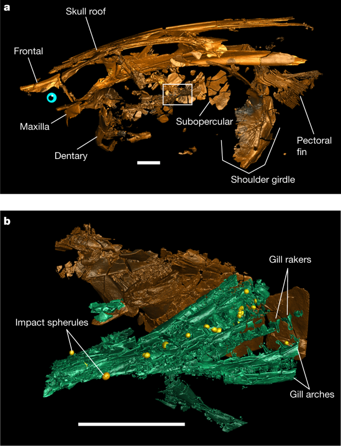
O depósito do evento Tanis em Dakota do Norte (EUA) é um depósito de seiche excepcional preservando um rico thanatocoenose (isto é, um conjunto de morte em massa) da mais recente biota cretácea no topo da Formação hell creek. A maioria dos macrofísis encontrados na localidade de Tanis representam baixas diretas do impacto do bólido K-Pg que foram enterrados dentro do depósito de seiche induzido pelo impacto5. Dezenas de minutos após o impacto, o seiche agitava grandes volumes de água e solo no estuário do rio Tanis5. As the seiche proceeded upstream, it advected bones, teeth, bivalves, ammonites, benthic foraminifera (Extended Data Fig. 1a–c) and plant matter in the suspended load while impact spherules rained down from the sky5. Within the thanatocoenotic accumulation, abundant acipenseriforms—sturgeons and paddlefishes—became oriented along the seiche flow directions and buried alive with numerous impact spherules in their gills5 (Fig. 1, Extended Data Fig. 2a, b).
a renderização tridimensional de fau de peixe-remo. DGS. ND.161.4559.T na vista lateral esquerda com a localização de uma varredura de maior resolução (retratada em b) indicada (contorno branco). b, renderização tridimensional do subopercular e brânquias em um com esferes de impacto preso (amarelo). Barras de escala, 2 cm. Dados tomográficos bidimensionais e renderizações tridimensionais totalmente anotadas são fornecidos em Extended Data Fig. 2. Uma renderização animada tridimensional da FAU. DGS. ND.161.4559.T é fornecido como Vídeo Suplementar 1.
Durante o Maastrichtian (ou seja, a última era do Cretáceo), o clima da atual Dakota do Norte envolveu quatro estações que foram documentadas em registros de anéis de árvores recuperados de outros locais do Alto Cretáceo na Formação hell creek12,13. Tanis foi localizado a aproximadamente 50° N durante o último Cretáceo e experimentou estancia distinta nas chuvas e temperatura14. As temperaturas regionais do ar foram reconstruídas para variar de 4 a 6 °C no inverno até uma média de cerca de 19 °C no verão13,14. Para descobrir a estação do impacto do bólido K-Pg, analisamos registros osteohistológicos de aposição óssea acipenseriform em três dentários de peixe-remo e três espinhas de barbatanas peitorais de esturjão que foram escavadas no sítio de Tanis em 2017 (Dados Estendidos Fig. 1d-j). Esses elementos esqueléticos preservam registros de crescimento não alterados do desenvolvimento embrionário até a morte, tornando-os altamente adequados para reconstruções da história da vida15,16.
Registros de crescimento de peixes cretáceos finais
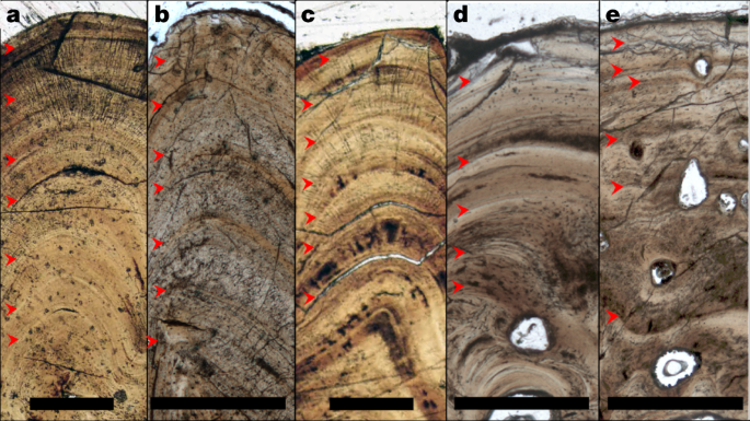
Para traçar o crescimento apposicional e identificar a estação em que a aposição óssea terminou, primeiro avaliamos a preservação dos padrões de crescimento ósseo em todos os espécimes estudados. Preparamos fatias ósseas dérmicas de seis espécimes acipenseriformes como lâminas microscópicas e as submetemos à avaliação osteohistológica, durante as quais linhas de crescimento preso (LAGs) foram facilmente reconhecidas (Fig. 2). Para corroborar a natureza anual dos LAGs usando osteohistologia virtual de alta resolução17,18, três dimensões (3D) volumes foram produzidos com a propagação de radiação síncrotron de contraste de fase microcomputada tomografia microcomputada19 na linha de feixe BM05 do European Synchrotron Radiation Facility, França. A natureza 3D dos dados síncrotrons permite a projeção ideal do padrão de deposição óssea em vários planos transversais e resolveu a relação exata entre sazonalidade e apposição óssea cíclica em detalhes soberbos20. In addition, virtual osteohistology allowed us to visualize the seasonal fluctuations of osteocyte lacunar density and volume, which are poorly expressed in the physical 2D thin sections18 (Fig. 3c, d) . The osteohistological data (Figs. 2, 3, Extended Data Figs. 3–6) were complemented with an incremental carbon isotope record extracted from one of the paddlefish dentaries (VUA.GG.2017.X-2724).
a–e, Thin sections in transmitted light of VUA.GG.2017.MDX-3 (a), VUA.GG.2017.X-2743M (b), VUA.GG.2017.X-2744M (c), VUA.GG.2017.X-2733A (d) and VUA.GG.2017.X-2733B (e), showing congruent pacing of bone apposition during the final years of life, terminating in spring. Red arrows indicate LAGs. Scale bars, 0.5 mm.
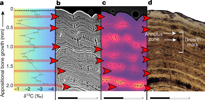
a, 𝛿13C record expressed as ‰ on the Vienna Pee Dee Belemnite (VPDB) reference scale. The colour gradient highlights the theoretical range between maximum values during seasonal (summer) trophic increase of 13C (yellow) and minimum values during trophic decrease of 13C (winter) (blue). b, Virtual thick section (average-value projection with 0.1-mm depth) showing growth zones during the favourable growth seasons and annuli and LAGs outside the favourable growth seasons. c, Cell density map51 of a virtual thick section (minimum-value projection with 0.2-mm depth) showing fluctuating osteocyte lacunar densities and sizes, with higher densities and largest sizes recorded during the favourable growth seasons (orange) and lower densities and smaller sizes outside the favourable growth seasons (purple). A comparative image of a larger section of bone with scale is provided in Extended Data Fig. 6. d, Microscopic thin section in transmitted light showing LAGs (red arrows) and a single growth mark indicated (bracket) spanning the distance between two subsequent LAGs and including a zone and an annulus (Extended Data Fig. 10b). Scanning data visualized in b and c were obtained approximately 10-mm distal from the physically sectioned thin slice of d, which itself was located directly proximal to the thick section sampled for a. Scale bars, 1 mm. Corresponding osteohistological data of the other five sampled acipenseriform fishes are presented in Extended Data Figs. 3–5.
The tomographic data show that impact spherules associated with the paddlefish skeleton are present exclusively in its gill rakers5 and are absent elsewhere in the preserved specimen (Fig. 1). The absence of impact spherules outside the gill rakers demonstrates that spherules were filtered out of the surrounding waters but had not yet proceeded into the oral cavity or further down the digestive tract, and had not impacted the fish carcases during perimortem exposure. Impact spherule accumulation in the gill rakers and the arrival of the seiche waves must therefore have occurred simultaneously5, which implies that the acipenseriforms were alive and foraging during the bolide impact and the last minutes of the Cretaceous.
Well-conserved bone growth archives
The degree of preservation of the sampled acipenseriform bones was assessed using micro-X-ray fluorescence (Methods, Extended Data Figs. 7–9), which would reveal potential taphonomic elemental exchange that may have affected the primary stable isotope composition. The micro-X-ray fluorescence maps show that Fe and Mn oxides are present in the bone vascular canals and surrounding sediments (Extended Data Fig. 8), but have not invaded the bone apatite (Ca5(PO4,CO3)3(OH,F,Cl)). Detrital components, characterized by high concentrations of K and Si, remain restricted to the sediment matrix (Extended Data Fig. 8f–j). The bone apatite conserves a highly homogeneous distribution of P and Ca (Extended Data Fig. 9), which corroborates the unaltered preservation of these apatitic tissues. Skeletal remains of the paddlefishes and sturgeons thus experienced negligible diagenetic alteration, probably as a consequence of rapid burial and possibly aided by early Mn and Fe oxide seam formation21,22. The exquisite 3D preservation of delicate structures, including non-ossified tissues that originally enveloped the brain (Extended Data Fig. 2c–f), further demonstrates the excellent preservation of the fossils and absence of taphonomic reorganization23.
Consistent records of a spring death
Paddlefish dentaries form through perichondral ossification around the Meckel’s cartilage24. Sturgeon pectoral fin spines consist of dermal bone—an intramembranous skeletal tissue that forms in the mesenchyme (mesodermal embryonic tissue)25. Unlike endochondral bone, perichondral and dermal bone do not originate through mineralization of cartilaginous precursors26,27,28 but grow exclusively through incremental bone matrix apposition by secretion of a row of osteoblasts24,26,27,28. The thickness of one annual growth mark cumulatively spans a thick (favourable) growth zone, a thinner (slowly deposited) annulus and, ultimately, a LAG20. Our microscopic and virtual osteohistological data consistently show that the six fishes perished (that is, stopped growing) while forming a growth zone shortly after a LAG was deposited (Figs. 2, 3, Extended Data Figs. 3–6), which coincides with an early stage of the favourable growth season20. The outermost cortices of all six acipenseriform individuals studied here also exhibit increasing osteocyte lacunar densities and sizes towards their periosteal surfaces (Fig. 3c, Extended Data Figs. 5, 6). In all specimens, this density remained lower than the highest densities and average sizes recorded in previous years (Fig. 3c, Extended Data Figs. 3–6, 10b). As osteocyte lacunar density and size patterns were consistently cyclical across the preceding years during which they peaked at the climaxes of the growth seasons, the last recorded growth season had thus not yet climaxed at the time of death (Figs. 2, 3, Extended Data Figs. 3–6, 10b).
The inferred annual growth cycles are independently corroborated by a stable carbon isotope (𝛿13Csc) archive that recorded several years of seasonal dietary fluctuations in growing bone. Paddlefish VUA.GG.2017.X-2724 also yielded, in addition to this 𝛿13Csc archive, an oxygen isotope (𝛿18Osc) record across the final six years of its life (Supplementary Data Table 1, Extended Data Fig. 10a, Methods). The low and constant 𝛿18Osc valores em VUA. GG.2017.X-2724 refletem a habitação exclusiva de ambientes de água doce pelos peixes-remo. Isso implica que seus registros osteohistológicos devem ter capturado a variabilidade sazonal em vez de, por exemplo, migração entre habitats salinos e de água doce. Embora os esturjões modernos sejam conhecidos por terem estilos de vida anadromos29,30, isso ainda está a ser confirmado para os esturjões fósseis em Tanis, pois os dados isotópicos das espinhas peitorais de esturjão não puderam ser protegidos (Métodos, 'Micromill'). Notavelmente, os registros osteohistológicos de todos os nossos esturjões e peixes-remo convergem para a mesma fase de crescimento anual, apesar de seus potenciais estilos de vida diferentes.
Como seus parentes modernos, os últimos peixes-remo maastrichtian de Tanis eram alimentadores de filtros que presumivelmente consumiam copépodes e outros zooplânctons29,30,31. Esses peixes provavelmente experimentaram um padrão anual de alimentação, determinado pela disponibilidade flutuante de alimentos, que atingiu o pico entre a primavera e o outono31. Durante a máxima produtividade, o zooplâncton ingerido enriquece o esqueleto crescente de peixes que alimentam filtros com 13C em relação a 12C32,33. Assim, o ciclicamente elevado 13C/12Relações C em peixe-remo VUA. GG.2017.X-2724 (Fig. 3a) refletem episódios distintos de alta disponibilidade e consumo de alimentos. Isótopos de carbono registram o registro de crescimento do Paddlefish VUA. GG.2017.X-2724 indicam que o pico de crescimento anual ainda não foi atingido e a temporada de alimentação ainda não havia clímax — corroborando uma morte na primavera boreal.
Implicações para a sobrevivência seletiva de K-Pg
O impacto do bólido de Chicxulub causou um pulso de calor global que desencadeou incêndios generalizados9,34. Após esta onda de calor, a última primavera boreal do Mesozoico passou para um inverno de impacto global10. Embora um calendário de junho para o impacto K-Pg tenha sido sugerido com base em indicações paleobotânicas para congelamento anômeo nesta região (Wyoming, EUA)35, as identidades paleobotânicas, inferências tafonômicas e suposições estratigráficas subjacentes a essa conclusão foram refutadas desde então.36,37,38,39. Além disso, o resfriamento pós-impacto aconteceu nos primeiros meses a décadas após o impacto k-pg10, o que torna os proxies registrando condições de congelamento pós-impacto assíncronsas com o próprio evento de impacto.
Um conjunto de fenômenos induzidos pelo impacto contribuiu para a extinção de K-Pg em diferentes escalas de tempo40,41. Nos dias seguintes ao impacto, seus efeitos instantâneos, como a radiação infravermelha intensa causada pela reentrada da ejeção34, resultando em incêndios florestais9,34 e a propagação de aerossóis sulfúricos levando à precipitação ácida42 deve ter afligido predominantemente os ambientes continentais expostos. Embora negociar essas condições hostis não tivesse garantido a sobrevivência, uma erradicação precoce de toda a vestia sempre significaria a extinção imediata41.
O timing sazonal do catastrófico impacto do boleto final-Cretáceo coloca o evento em um estágio particularmente sensível para ciclos de vida biológico no Hemisfério Norte. Em muitos impostos, a reprodução anual e o crescimento ocorrem durante a primavera. Espécies com tempos de incubação mais longos, como répteis não-aviários, incluindo pterossauros e a maioria dos dinossauros, eram indiscutivelmente mais vulneráveis a perturbações ambientais repentinas do que outros grupos43 (por exemplo, pássaros). Os ecossistemas do Hemisfério Sul, que foram atingidos durante o outono austral, parecem ter se recuperado até duas vezes mais rápido que as comunidades do Hemisfério Norte44, consistente com um efeito sazonal na recuperação biótica.
O acolhimento subterrâneo, concebivelmente, contribuiu para a sobrevivência cinodonte da crise permo-triássica (PT)45. Da mesma forma, incêndios em larga escala que se espalham pelo Hemisfério Sul9,34,41 pode ter sido evitado por mamíferos hibernando que já estavam abrigados em tocas34,41 em antecipação ao inverno austral. Modos adicionais de dormência sazonal, torpor e/ou estivação, que hoje são praticados por vários mamíferos46,47 as well as certain amphibians, birds and crocodilians48, could have facilitated further underground survival. In the aftermath of the K–Pg event, ecological networks collapsed from the bottom up. Floral necrosis9 and extinction immediately affected species dependent on primary producers, while some animals capable of exploiting alternative resources—for example, certain birds and mammals49,50—persisted.
Conclusions
Seasonal timing of the Chicxulub impact in boreal spring and austral autumn will aid in further calibrating evolutionary models exploring the selectivity of the K–Pg extinction and the asymmetry in extinction and recovery patterns between the two hemispheres. Decoupling short- and long-term effects of the bolide impact on the K–Pg mass extinction will also aid in identifying extinction risks and modes of ecological deterioration caused by the forthcoming global climate change. The uniquely constrained Tanis site5 offers valuable proxies for reconstructing the environmental, climatological and biological conditions that prevailed locally when the Mesozoic ended.
Methods
Fieldwork
Excavation at the Tanis locality in south-western North Dakota took place between 10 August and 20 August 2017. Sections of dentaries of paddlefishes and pectoral fin spines of sturgeons were collected in the field for histological study.
Thin sectioning
Four out of the six samples were excavated from the sediment matrix. These included all sturgeon pectoral fin spines (VUA.GG.2017.X-2743M, VUA.GG.2017.X-2744M, and VUA.GG.2017.MDX-3) and one of the paddlefish dentaries (VUA.GG.2017.X-2724). Paddlefish dentaries VUA.GG.2017.X-2733A and VUA.GG.2017.X-2733B, belong to two individuals, were uncovered aligned to each other and fractured upon discovery. To avoid further damage, the samples were embedded in epoxy resin prior to thin sectioning. All specimens were cut with a diamond saw and polished to obtain microscopic thin sections (about 50-μm thick) and thick sections for micromilling (about 200-μm thick). See Extended Data Fig. 1e–j for images of the specimens and the sampling locations.
Osteohistological analysis
In the acipenseriform dermal bones examined in this study, annual growth cyclicity can be traced through growth marks (GMs).
A GM spans a single growth cycle that typically lasts one year and can be divided into a zone, an annulus, and a LAG20,52. The zone is deposited during a period of relative rapid growth in the active or favourable growth season20. The annulus is subsequently formed when growth slows down towards the end of the growth season20. Finally, a LAG forms when growth periodically ceases until the next growth season starts and a new zone is deposited20.
During the formation of a growth zone, the density and volumes of osteocyte lacunae (OL; subcircular dark features in Extended Data Fig. 10a) initially increase when growth accelerates. Subsequently, towards and into the annulus, OL density and volume decrease as growth slows down18. Because a LAG coincides with a temporary arrest of local osteogenesis, it is only expressed when deposition of a new growth zone has commenced. All six studied specimens show a LAG relatively close to the outermost partial growth zone.
In fossil bone, LAGs often appear as sharply defined dark lines53 that typically constitute a poorly coherent interface between adjacent bone layers, thus facilitating (local) delamination between adjacent cortical layers53. During fossilization, percolation products can accumulate in these gaps and thereby (locally) accentuate the LAGs51,53 (figure 31.3G of ref. 52). Based on this well-understood expression of LAGs (that we recognize from our own experience as well; S.S. personal observation), we have consistently identified the LAGs as locally stained dark lines that may be associated with circumferentially propagated cracked surfaces which are oriented parallel to the periosteal deposits.
Besides cyclical seasonal factors that synchronize GM accretion, stress may induce additional diapause stages that result in supplementary marks within a single year54. Cessation of growth for the duration of several weeks can provoke the formation of a LAG54. However, such non-cyclical marks “tend to be haphazard rather than regular (that is, they do not reflect a particular spacing or rhythm)” and do not encircle the cortex of the skeletal element but “tend to be locally confined to an arc”55.
As the studied bones yield only regularly spaced GMs along their complete circumference, we confidently identify the preserved GMs as annual cycles. Moreover, the fluctuating quantified density and volumes of osteocyte lacunae (Extended Data Fig. 6d–f) and the carbon isotopic record (Fig. 3a, Extended Data Fig. 10a) across the final seven years of growth of VUA.GG.2017.X-2724 are exclusively consistent with the identification of annual LAGs in corresponding physical thin sections. In all studied specimens, bone growth terminated during the process of zonal bone growth.
Micro-X-ray fluorescence
Fragments of the paddlefish and sturgeon samples that remained after thin sectioning were analysed with microX-ray fluorescence. High-resolution elemental mapping was conducted using a Bruker M4 Tornado 2D spectrometer at 50 kV and 600 μA, without a filter, and at an acquisition rate of 20 μm per 5 ms at the Vrije Universiteit Brussel.
Micromill
The growth increments were sampled in the thick sections (about 200-μm thick) at the highest possible accuracy using a Micromill (Merkantek). Drill transects were assigned in the accompanying software and after each individual sample was collected, the drill bit was cleaned with ethanol. Not all thick sections were suitable for micromilling. The lobed anatomy of the sturgeon fin spines (VUA.GG.2017.X-2743M and VUA.GG.2017.X-2744M) proved too complex to reliably sample single growth increments with the micromill. Paddlefish dentaries VUA.GG.2017.X-2733A and VUA.GG.2017.X-2733B only exposed a few growth lines that were too narrow to sample with the micromill. Sturgeon pectoral fin spine VUA.GG.2017.MDX-3 and paddlefish dentary VUA.GG.2017.X-2724 were sampled up to the outermost growth increment.
Stable isotope analysis
Micromilled hydroxyapatite samples of specimen VUA.GG.2017.X-2724 weighing about 50 μg were placed in Exetainer vials (Labco) and flushed with purified helium gas. For reference, the analysed amounts of structural carbonate are equivalent to anout 5 μg of CaCO3. Orthophosphoric acid was subsequently added and allowed to react for 24 h at 45 °C. VUA.GG.2017.MDX-3 was routinely analysed with a Thermo Finnigan Deltaplus mass spectrometer connected to a Thermo Finnigan GasBench II at the Earth Sciences Stable Isotope Laboratory (Vrije Universiteit, Amsterdam). However, the amount of CO2 generated was found to be too small to permit reliable isotopic determinations. To alleviate this, the GasBench was provisionally interfaced with a cold trap in which the CO2 was frozen with liquid nitrogen during a 2 min period. After trapping for 2 min, an accurate single-pulse measurement was performed for each of the apatitic samples and standards. Each isotopic sample determination was preceded by six pulses of monitoring CO2 with a calibrated isotopic composition to assure stable conditions of the mass spectrometer. The isotopic measurements of the weighted micromilled samples were bracketed by the analyses of the inter-laboratorial apatite standard (Ag-Lox) to account for the linearity effect56. After corrections, the uncertainties for 𝛿13C and 𝛿18O of the Ag-Lox (n = 4) were 0.16 ‰ and 0.39 ‰ (1 s.d.) respectively. Although the amount of extracted and analysed structural carbonate remains insufficient for optimal isotopic determination, the relatively large recovered 𝛿13C variability still yields a meaningful record across the appositional bone archive. The 𝛿18O values of structural carbonate, unlike those of phosphate (PO4)57, do not offer a sensitive palaeo-environmental proxy for accurate seasonal temperature reconstructions58. However, the relatively constant 𝛿18O values of structural carbonate precludes large 𝛿18O changes in ambient water, such as shifts between freshwater and saline environments.
Propagation phase-contrast synchrotron radiation micro-computed tomography
Paddlefish specimen FAU.DGS.ND.161.4559.T lacks the paddle-shaped rostrum and all aspects caudal to the pectoral girdle. FAU.DGS.ND.161.4559.T was provided by the Palm Beach Museum of Natural History. Data acquisition took place in May 2018 on Beamline BM05 of the European Synchrotron Radiation Facility, Grenoble, France59. The complete specimen was scanned at an average energy of 132 keV using the white beam of BM05 filtered with 0.4 mm of Mo and 9 mm of Cu. The detector was composed of a 2-mm-thick LuAG:Ce scintillator optically coupled to a PCO edge 4.2 CLHS sCMOS camera. The resulting voxel size was 43.5 µm. To obtain sufficient propagation phasecontrast, the distance between the sample and the detector was set at 5 m. A total of 205 scans, each consisting of 5,000 projections taken at 7-ms intervals, were performed with a vertical displacement of 1.4 mm at a vertical field of view of 2.8 mm to ensure a double scan of the complete samples. Scans were performed in half-acquisition mode to enlarge the lateral field of view. The volume was reconstructed using a single-distance phase retrieval algorithm coupled with filtered back projection as implemented in the ESRF software PyHST2. Vertical concatenation, 16-bit conversion, and ring artefact corrections were performed using MATLAB scripts developed in-house. The gill region and impact spherules were subsequently scanned at a voxel size of 13.67 μm (filters: 0.4 mm of Mo and 6 mm of Cu, scintillator: LuAG:Ce, 500-μm thick, detected energy: 166 keV, propagation distance: 2.5 m). The samples were scanned in half-acquisition mode in two columns of 77 scans, each consisting of 4,998 projections with exposure times of 0.05 s, that were laterally concatenated after reconstruction. Finally, sample (VUA.GG.2017.X-2724) from the paddlefish dentaries and (VUA.GG.2017.MDX-3, VUA.GG.2017.X-2743M and VUA.GG.2017.X-2744M) of the sturgeon pectoral fin spines were scanned at 4.35 µm voxel size for osteohistological analysis60 (filters: 3.5 mm of Al plus 11 bars Al with a diameter of 5 mm, scintillator: LuAG:Ce scintillator, 500-µm thick, detected energy: 92 keV, propagation distance: 1.5 m). The samples were scanned in half-acquisition mode in one single column of 22 scans, each consisting of 4,998 projections with exposure times of 60 ms.
Digital 3D extraction of the bones and impact spherules was performed in VGStudio MAX 3.2 (Volume Graphics). VGStudio MAX 3.2 furthermore enabled the creation of virtual thick sections of the osteohistological samples through the ‘thick slab-mode’, which captures the maximum, average, or minimum, grey-level values along the desired field depth. Virtual thick sections were obtained from the average grey-level values at a thickness of 100 µm following optimal 3D alignment of the annuli and LAGs. Additional virtual thick sections were created from the minimum grey-level values at a thickness of 200 µm to best resolve the sizes and distributions of osteocyte lacunae. A coloured map of the density of the osteocyte lacunar distribution was created with a Gaussian filter51. Finally, we visualized the annual cyclicity of osteocyte lacunar volumes18 in paddlefish dentary VUA.GG.2017.X-2724. As the resolution of our data (voxel size of 4.35 μm; appropriate for assessing GMs and osteocyte lacunar distributions) is sixfold lower than that used for earlier osteocyte lacunar volumetric quantification in fish bones18 (voxel size of 0.7 μm), our result should be considered with appropriate care. Closely spaced (large) osteocyte lacunae may occasionally be conjoined and additional phenomena in the broad size range of osteocyte lacunae may be incidentally included in the visualized distribution. Moreover, in tomographic data, osteocyte lacunae are delimited by slight colour gradients (rather than discrete lines) that scale with voxel size. Because the outermost feature fringe contributes disproportionally to recovered volumes, these values are somewhat skewed relative to the original osteocyte lacunar volumes, which likely produces exaggerated volume values. Therefore, although all rendered features were extracted with a single thresholding operation and relative patterns are conservatively retained, absolute volume values are best considered in a comparative context.
Reporting summary
Further information on research design is available in the Nature Research Reporting Summary linked to this paper.
Data availability
All isotopic, geochemical, and osteohistological data are included in the paper and Extended Data. Tomographic data of FAU.DGS.ND.161.4559.T, VUA.GG.2017.X-2724, VUA.GG.2017.MDX-3, VUA.GG.2017.X-2743M, and VUA.GG.2017.X-2744M are available at https://doi.org/10.5281/zenodo.5776294 and the http://paleo.esrf.eu database.
References
Alvarez, L. W., Alvarez, W., Asaro, F. & Michel, H. V. Extraterrestrial cause for the Cretaceous–Tertiary extinction. Science 208, 1095–1108 (1980).
Smit, J. & Hertogen, J. An extraterrestrial event at the Cretaceous–Tertiary boundary. Nature 285, 198–200 (1980).
Raup, D. M. Biological extinction in earth history. Science 231, 1528–1533 (1986).
Schulte, P. et al. The Chicxulub asteroid impact and mass extinction at the Cretaceous–Paleogene boundary. Science 327, 1214–1218 (2010).
DePalma, R. A. et al. A seismically induced onshore surge deposit at the KPg boundary, North Dakota. Proc. Natl Acad. Sci. USA 116, 8190–8199 (2019).
Smit, J. et al. Tektite-bearing, deep-water clastic unit at the Cretaceous–Tertiary boundary in northeastern Mexico. Geology 20, 99–103 (1992).
Alvarez, W. in The Cretaceous-Tertiary Event and Other Catastrophes in Earth History (eds Ryder, G. et al.) 141–150 (Geological Society of America, 1996).
Smit, J. The global stratigraphy of the Cretaceous–Tertiary boundary impact ejecta. Annu. Rev. Earth Planet. Sci. 27, 75–113 (1999).
Morgan, J., Artemieva, N. & Goldin, T. Revisiting wildfires at the K–Pg boundary. J. Geophys. Res. 118, 1508–1520 (2013).
Vellekoop, J. et al. Rapid short-term cooling following the Chicxulub impact at the Cretaceous–Paleogene boundary. Proc. Natl Acad. Sci. USA 111, 7537–7541 (2014).
Vellekoop, J. et al. Evidence for Cretaceous-Paleogene boundary bolide ‘impact winter’ conditions from New Jersey, USA. Geology 44, 619–622 (2016).
Golovneva, L. B. The Maastrichtian (Late Cretaceous) climate in the northern hemisphere. J. Geol. Soc. Lond. 181, 43–54 (2000).
Wolfe, J. A. & Upchurch Jr, G. R. North American nonmarine climates and vegetation during the Late Cretaceous. Palaeogeogr. Palaeocl. 61, 33–77 (1987).
Hallam, A. A review of Mesozoic climates. J. Geol. Soc. London 142, 433–445 (1985).
Adams, L. A. Age determination and rate of growth in Polyodon spathula, by means of the growth rings of the otoliths and dentary bone. Am. Midl. Nat. 28, 617–630 (1942).
Bakhshalizadeh, S., Bani, A., Abdolmalaki, S. & Moltschaniwskyj, N. Identifying major events in two sturgeons’ life using pectoral fin spine ring structure: exploring the use of a non-destructive method. Environ. Sci. Pollut. R 24, 18554–18562 (2017).
Sanchez, S., Ahlberg, P. E., Trinajstic, K. M., Mirone, A. & Tafforeau, P. Three-dimensional synchrotron virtual paleohistology: a new insight into the world of fossil bone microstructures. Microsc. Microanal. 18, 1095–1105 (2012).
Davesne, D., Schmitt, A. D., Fernandez, V., Benson, R. B. & Sanchez, S. Three‐dimensional characterization of osteocyte volumes at multiple scales, and its relationship with bone biology and genome evolution in ray‐finned fishes. J. Evol. Biol. 33, 808–830 (2020).
Tafforeau, P., Bentaleb, I., Jaeger, J.-J. & Martin, C. Nature of enamel laminations and mineralization in rhinoceros enamel using histology and X-ray synchrotron microtomography: potential implications for palaeoenvironmental isotopic studies. Palaeogeogr. Palaeocl. 246, 206–227 (2007).
Castanet, J. Bone—Volume 7: Bone Growth (ed. Hall, B. K.) 245–283 (CRC Press, 1993).
Hedges, R. E. Bone diagenesis: an overview of processes. Archaeometry 44, 319–328 (2002).
Dumont, M. et al. Synchrotron XRF analyses of element distribution in fossilized sauropod dinosaur bones. Powder Diffr. 24, 130–134 (2009).
Pradel, A. et al. Skull and brain of a 300-million-year-old chimaeroid fish revealed by synchrotron holotomography. Proc. Natl Acad. Sci. USA 106, 5224–5228 (2009).
Weigele, J. & Franz‐Odendaal, T. A. Functional bone histology of zebrafish reveals two types of endochondral ossification, different types of osteoblast clusters and a new bone type. J. Anat. 229, 92–103 (2016).
Enlow, D. H. The Human Face. An Account of the Postnatal Growth and Development of the Craniofacial Skeleton (Harper and Row, 1968).
Grande, L. & Bemis, W. E. Osteology and phylogenetic relationships of fossil and recent paddlefishes (Polyodontidae) with comments on the interrelationships of Acipenseriformes. J. Vert. Paleo. 11, 1–121 (1991).
De Ricqlès, A. J., Meunier, F. J., Castanet, J. & Francillon-Vieillot, H. Bone 3, Bone Matrix and Bone Specific Products (CRC Press, 1991).
Hall, B. K. Bones and Cartilage: Developmental and Evolutionary Skeletal Biology (Elsevier, 2005).
Bemis, W. E. & Kynard, B. Sturgeon rivers: an introduction to acipenseriform biogeography and life history. Environ. Biol. Fish 48, 167–183 (1997).
LeBreton, G. T., Beamish, F. W. H. & McKinley, S. R. (eds) Sturgeons and paddlefish of North America, Vol. 27 (Springer, 2004)
Blackwell, B.G., Murphy, B. R. & Pitman, V.M. Adequação de recursos alimentares e parâmetros físico-químicos no baixo Rio Trinity, Texas para peixe-remo. Freshw. O Ecol. 10, 163-175 (1995).
Fry, B. & Sherr, E.B. δ13Medidas C como indicadores de fluxo de carbono em ecossistemas marinhos e de água doce. Stud. https://doi.org/10.1007/978-1-4612-3498-2_12 (1989).
Finlay, J.C. Relação estável‐isótopo de carbono da biota do rio: implicações para o fluxo de energia em teias de alimentos loticos. Ecologia 82, 1052-1064 (2001).
Robertson, D.S., Lewis, W.M., Sheehan, P.M. & Toon, O.B. K-Pg extinction: reavaliação da hipótese de calor-fogo. Geofisias. Res. 118, 329-336 (2013).
Wolfe, J. A. Palaeobotanical evidência para um "inverno de impacto" de junho no limite Cretáceo/Terciário. Natureza 352, 420-423 (1991).
Nichols, D.J. Plants na fronteira K/T. Natureza 356, 295-295 (1992).
Hickey, L. J. & McWeeney, L. J. Plants na fronteira K/T. Natureza 356, 295-296 (1992).
McIver, E. E. Paleobotânica evidências para a ruptura do ecossistema na fronteira Cretáceo-Terciária de Wood Mountain, Saskatchewan, Canadá. Pode. J. Earth Sci. 36, 775-789 (1999).
Upchurch, G. R., Lomax, B. H. & Beerling, D. J. Paleobotânicas evidências de mudança climática através da fronteira Cretáceo-Terciária, América do Norte: vinte anos depois de Wolfe e Upchurch. O Cour. Forsch, o que está com o Forsch. Senck 258, 57 (2007).
Kring, D.A. O evento de impacto chicxulub e suas consequências ambientais no limite Cretáceo-Terciário. Palaeogeogr. O Palaeocl. 255, 4-21 (2007).
Robertson, D.S., McKenna, M.C., Toon, O.B., Hope, S. & Lillegraven, J. A. Survival nas primeiras horas do Cenozoico. Geol. Soc. Am. Bull. 116, 760-768 (2004).
D'Hondt, S., Pilson, M. E., Sigurdsson, H., Hanson Jr, A. K. & Carey, S. Surface-water acidification and extinction at the Cretaceous-Tertiary boundary. Geologia 22, 983-986 (1994).
Erickson, G.M., Zelenitsky, D.K., Kay, D. I. & Norell, M. A. Períodos de incubação de dinossauros diretamente determinados a partir da contagem de linhas de crescimento em dentes embrionários mostram desenvolvimento de grau réptil. Proc. Natl Acad. Sci. USA 114, 540-545 (2017).
Donovan, M. P., Iglesias, A., Wilf, P., Labandeira, C.C. & Cúneo, N. R. Rápida recuperação das associações patagônicas de plantas-insetos após a extinção do Cretáceo final. Nat. Ecol. Evol. 1.0012 (2016).
Fernandez, V. et al. Síncrotron revela casal estranho triássico: anfíbio ferido e aestivação de torção de táróide. PLoS ONE 8, e64978 (2013).
Nowack, J., Cooper, C. E. & Geiser, F. Cool echidnas sobrevivem ao incêndio. Proc. R. Soc.B 283, 20160382 (2016).
Lovegrove, B. G., Lobban, K. D. & Levesque, D. L. Mamíferos sobrevivência na fronteira Cretáceo-Palaeogene: homeostase metabólica em hibernação tropical prolongada em tenrecs. Proc. R. Soc.B 281, 20141304 (2014).
Withers, P.C. & Cooper, C. in Enciclopédia de Ecologia (eds Jorgensen, S. E. & Fath, B.) 952-957 (Elsevier, 2008).
Field, D.J. et al. Evolução precoce das aves modernas estruturadas pelo colapso florestal global no final da extinção em massa do Cretáceo. Curr. Biol. 28, 1825-1831 (2018).
Schleuning, M. et al. As redes ecológicas são mais sensíveis à vegetação do que à extinção animal sob as mudanças climáticas. Nat. Commun. 7, 13965 (2016).
Sanchez, S. et al.3D arquitetura microestrutural de apegos musculares em vertebrados antigos e fósseis revelados pela microtomografia síncrotron. Plos ONE 8, e56992 (2013).
de Buffrénil, V., Quilhac, A. & Castanet, J. in Vertebrate Skeletal Histology and Paleohistology (eds de Buffrénil, V. et al.) 626-645 (Routledge, 2021).
Lee, A. H. & O'Connor, P.M. A histologia óssea confirma o crescimento determinante e o pequeno tamanho do corpo no terópode noasúrido Masiakasaurus knopfleri. J. Vertebr. Paleontol. 33, 865-876 (2013).
Klevezal, G. A. & Stewart, B. S. Patterns e calibração de camadas em cimento dente de focas-elefante fêmeas do norte, Mirounga angustirostris. Mamífero. 75, 483-487 (1994).
Woodward, H.N., Padian, K. e Lee, A.H. Em Histologia Óssea de Tetrápodes Fósseis — Métodos avançados, Análise e Interpretação (eds Padian, K. & E. T. Lamm) 195-215 (Univ. California Press, 2013).
Vonhof, H.B. et al. Análise de isótopos estáveis de alta precisão de < caCO de 5 μg3 amostras por espectrometria de massa de fluxo contínuo. Comuna rápida. Mass Spectr. 34, e8878 (2020).
Pucéat, E. et al. Equação de fracionamento fosfato-água revisada reavaliando paleotemperaturas derivadas de apatita biogênica. Planeta Terra. Sci. Lett. 298, 135-142 (2010).
Vennemann, T. W., Hegner, E., Cliff, G. & Benz, G. W. Composição isotópica dos recentes dentes de tubarão como um proxy para as condições ambientais. O Geochim. A Cosmochim. Acta 65, 1583-1599 (2001).
Tafforeau, P. et al. Applications of X-ray synchrotron microtomography for non-destructive 3D studies of paleontological specimens. Appl. Phys. A 83, 195–202 (2006).
Tafforeau, P. & Smith, T. M. Nondestructive imaging of hominoid dental microstructure using phase contrast X-ray synchrotron microtomography. J. Hum. Evol. 54, 272–278 (2008).
Acknowledgements
M.A.D.D. was partially funded by an EAVP Research Grant (ERG) awarded by the European Association of Vertebrate Palaeontologists. D.F.A.E.V. gratefully acknowledges support from the Wenner-Gren Foundation through fellowships UPD2018-250 and UPD2019-0076. VGStudio Max (Volume Graphics, Germany) and the Porosity/Inclusion Analysis module were funded by the Vetenskapsrådet through grants 2015-04335; 2019-04595 to S.S. We thank R. DePalma for providing guidance in the field and access to the specimens. We acknowledge the ESRF for provisioning beamtime at BM05. We thank V. Fernandez and K. Chapelle for their assistance with the segmentation in VGStudio; B. Lacet for help with the preparation of the thin and thick sections; M. Hagen for the use of her sedimentology laboratory and the microbalance for weeks in a row; F. Peeters for assistance in photographing the thin sections while sharing his thoughts on the project; and P. Ahlberg for his advice, labelling of the paddlefish bones, fruitful discussions and invaluable consultation.
Funding
Open access funding provided by Uppsala University.
Author information
Affiliations
Contribuições
M.A.D.D., J.S. e H.J.L.v.d.L. conceberam e projetaram o projeto. Os materiais foram escavados pela M.A.D.D. em 2017. M.A.D.D., D.F.A.E.V., C.B. e P.T. realizaram os experimentos síncrotrons. K.H.W.S. e M.A.D.D. realizaram a análise de fluorescência de micro raios-X. M.A.D.D. amostrado os espécimes com a microimilia. M.A.D.D., S.J.A.V.-W. e H.J.L.v.d.L realizaram as análises de isótopos. P.T. processou e reconstruiu a fase de propagação bruta contraste dados de tomografia micro computada de radiação. M.A.D.D. e D.F.A.E.V. segmentaram os dados de digitalização. M.A.D.D., J.S., D.F.A.E.V., S.S. e H.J.L.v.d.L. analisaram os dados. A S.S. criou a Fig. 3c e a Extended Data Fig. 8a-c. D.F.A.E.V. criou o Extended Data Fig. 8d–f. M.A.D.D. criou todas as outras figuras. Todos os autores discutiram as interpretações. M.A.D.D., D.F.A.E.V. e H.J.L.v.d.L. escreveram o manuscrito. Todos os autores forneceram uma revisão crítica e aprovaram a redação final do manuscrito.


Nenhum comentário:
Postar um comentário
Observação: somente um membro deste blog pode postar um comentário.