quinta-feira, 31 de maio de 2018

The origin of squamates revealed by a Middle Triassic lizard from the Italian Alps

Naturevolume 557pages706709 (2018) | Download Citation

Abstract

Modern squamates (lizards, snakes and amphisbaenians) are the world’s most diverse group of tetrapods along with birds1 and have a long evolutionary history, with the oldest known fossils dating from the Middle Jurassic period—168 million years ago2,3,4.

The evolutionary origin of squamates is contentious because of several issues: (1) a fossil gap of approximately 70 million years exists between the oldest known fossils and their estimated origin5,6,7; (2) limited sampling of squamates in reptile phylogenies; and (3) conflicts between morphological and molecular hypotheses regarding the origin of crown squamates6,8,9. Here we shed light on these problems by using high-resolution microfocus X-ray computed tomography data from the articulated fossil reptile Megachirella wachtleri (Middle Triassic period, Italian Alps10).

We also present a phylogenetic dataset, combining fossils and extant taxa, and morphological and molecular data. We analysed this dataset under different optimality criteria to assess diapsid reptile relationships and the origins of squamates. Our results re-shape the diapsid phylogeny and present evidence that M. wachtleri is the oldest known stem squamate. Megachirella is 75 million years older than the previously known oldest squamate fossils, partially filling the fossil gap in the origin of lizards, and indicates a more gradual acquisition of squamatan features in diapsid evolution than previously thought.

For the first time, to our knowledge, morphological and molecular data are in agreement regarding early squamate evolution, with geckoes—and not iguanians—as the earliest crown clade squamates. Divergence time estimates using relaxed combined morphological and molecular clocks show that lepidosaurs and most other diapsids originated before the Permian/Triassic extinction event, indicating that the Triassic was a period of radiation, not origin, for several diapsid lineages.

Euryhaline ecology of early tetrapods revealed by stable isotopes

Ecologia de Euryhalina dos primeiros tetrápodes revelada por isótopos estáveis

Nature (2018) | Download Citation

Abstract

The fish-to-tetrapod transition—followed later by terrestrialization—represented a major step in vertebrate evolution that gave rise to a successful clade that today contains more than 30,000 tetrapod species. The early tetrapod Ichthyostega was discovered in 1929 in the Devonian Old Red Sandstone sediments of East Greenland (dated to approximately 365 million years ago). Since then, our understanding of the fish-to-tetrapod transition has increased considerably, owing to the discovery of additional Devonian taxa that represent early tetrapods or groups evolutionarily close to them. However, the aquatic environment of early tetrapods and the vertebrate fauna associated with them has remained elusive and highly debated. Here we use a multi-stable isotope approach (δ13C, δ18O and δ34S) to show that some Devonian vertebrates, including early tetrapods, were euryhaline and inhabited transitional aquatic environments subject to high-magnitude, rapid changes in salinity, such as estuaries or deltas. Euryhalinity may have predisposed the early tetrapod clade to be able to survive Late Devonian biotic crises and then successfully colonize terrestrial environments.

quarta-feira, 30 de maio de 2018

160-Million-Year-Old Pterosaur Ate Like a Flamingo


Partner Series
160-Million-Year-Old Pterosaur Ate Like a Flamingo
The skull of the newfound species Liaodactylus primus, the earliest filter-feeding pterosaur on record.
Credit: Chang-Fu Zhou
During the dinosaur era, pterosaurs would swoop down and snap up wriggly fish and buzzing insects with their spiky teeth, with the exception of one odd group — pterosaurs that ate their meals like modern-day flamingos do: by filter feeding.

Now, researchers have found the earliest filter-feeding pterosaur on record. The specimen, which was discovered in northeast China's Liaoning province, is 160 million years old, and dates to the Jurassic period (199.6 million to 145.5 million years ago), according to a new study.
"The fossil specimen represents a medium-sized pterosaur with a large number of fine teeth indicative of filter-feeding adaptation," said the study's co-researcher Ke-Qin Gao, a professor at the School of Earth and Space Sciences at Peking University in China. [Photos of Pterosaurs: Flight in the Age of Dinosaurs]

Researchers named the newly identified pterosaur Liaodactylus primus — honoring the Liaoning province by combining it with the suffix "dactylos," the Greek word for "finger," or "winged finger," which is commonly used for pterosaur names. The species name "primus" is Latin for "first," an indication of the pterosaur's early age in the pterodactyloid group, the researchers said.
The pterosaur's jaw was almost 50 percent of its roughly 5-inch-long (13.3 centimeter) skull, the researchers said. Its wingspan was likely about 3.2 feet (1 meter), or slightly smaller than the wingspan of a great horned owl (Bubo virginianus), Gao said.

Despite its small size, L. primus is a big finding, because it helps researchers learn more about when filter-feeding pterosaurs developed, Gao said.
"The new pterosaur marks a major ecological transition in pterosaur evolution from fish-catching or insect-eating to filter-feeding adaptation," Gao told Live Science in an email. "This critical transition was followed by a burst of ecological diversification of pterosaurs, which had a significant impact on the change of the terrestrial ecosystems of the Cretaceous world."
The newfound pterosaur species was uncovered at the Daxishan site in China's Liaoning province, an area famous for its well-preserved fossils of dinosaurs with feathers. The pterosaur was discovered in rock that's about 160 million years old.
Credit: Zhou C.F. et al. Royal Society Open Science (2017)
Although pterosaurs lived during the dinosaur age, known as the Mesozoic era, they were not dinosaurs themselves. Rather, pterosaurs were flying reptiles — the first flying vertebrates on record, the researchers said.

Pterosaurs initially appeared about 215 million years ago during the Triassic period, and went extinct when a 6-mile-wide (10 kilometer) asteroid slammed into Earth about 65 million years ago, ending the Cretaceous period. The first known pterosaur was discovered in the 1760s in Germany, and now there are more than 110 pterosaur species known worldwide, the researchers wrote in the study, including a pterosaur with 110 teeth that was unearthed in Utah and a cat-size pterosaur discovered in British Columbia, Canada.

All of these pterosaurs ate a variety of different meals. During the Cretaceous, pterosaurs "engaged in a variety of feeding adaptations, including filter-feeding, fish-eating, carnivory and scavenging, herbivory including frugivory, mollusk shell-crushing and omnivory," the researchers wrote in the study.
The new study was published online Wednesday (Feb. 1) in the journal Royal Society Open Science.
Original article on Live Science.

World's Largest Pterosaur Jawbone Discovered in Transylvania


World's Largest Pterosaur Jawbone Discovered in Transylvania
The reconstructed skull of Dracula, another pterosaur found in the same region of Romania as the newly analyzed specimen.
Credit: Axel Schmidt/Dinosaurier Museum
The largest pterosaur jawbone on record has just been analyzed, and it's so big that it likely helped the prehistoric beast gulp down freshwater turtles and large dinosaur eggs for dinner more than 66 million years ago, a new study finds.

The fossil of the pterosaur's robust lower jaw is a mere 7.4 inches (18.8 centimeters) long, but the jawbone likely measured longer than a yardstick — or between 37 and 43 inches (94 and 110 cm) — when the reptile was alive, the researchers wrote in the study.

This absurdly long jaw is "more than three times the size of the complete, 290-millimeter-long [11.4 inches] holotype mandible of Bakonydraco," a pterosaur that appears to be closely related to the newly analyzed creature, the researchers wrote in the study. [Photos: Baby Pterosaurs Couldn't Fly as Hatchlings]

Study co-researcher Dan Grigorescu, a geologist at the University of Bucharest in Romania, collected the fossilized jawbone at the junction of two creeks in the Hațeg Basin, near the village of Vặlioara, which is in Transylvania, Romania, in 1984. But the fossil wasn't recognized as belonging to a pterosaur until 2011, when lead study researcher Mátyás Vremir, a geologist at the Transylvanian Museum Society, and study co-researcher Gareth Dyke, a paleontologist at the University of Debrecen in Hungary, realized its importance, according to National Geographic.
The rocks where scientists found the pterosaur Dracula. The newly studied fossil comes from the same region in Romania.
Credit: Mátyás Vremir
During the Cretaceous period, when this pterosaur was alive, Hațeg Basin was an island inhabited by dwarf dinosaurs, which were smaller than their counterparts on the mainland. Vremir unearthed the fossilized remains of one of these weird, stocky dinosaurs — a predator known as Balaur bondoc — in 2009, Live Science previously reported.

But Hațeg is also known for large pterosaurs, including Hatzegopteryx, which likely stood as tall as a giraffe, with a wingspan of up to 36 feet (10.9 meters). Another pterosaur from Hațeg, nicknamed Dracula, had an even larger wingspan of up to 39 feet (12 m).

"Islands are notorious for throwing up oddities. We have a bunch of weird dinosaurs from Hațeg and a lack of really big carnivores, so the pterosaurs were basically tyrannosaur surrogates," Dave Hone, a paleontologist at Queen Mary University of London in England, told National Geographic.
The newly studied specimen is slightly smaller than Dracula, shown here.
Credit: Dinosaurier Museum
But just because the newly studied pterosaur — which has yet to be scientifically named — has the largest jawbone ever found, it doesn't necessarily mean it was the biggest pterosaur on record, the researchers said. Rather, it probably had a wingspan of over 26 feet (8 m) and likely belonged to a family of pterosaurs known as the Azhdarchids, the researchers wrote in the study.

"It's always exciting to see new Azhdarchid material in the literature, especially fossils of giant pterosaurs," Kierstin Rosenbach, a doctoral student in the Department of Earth and Environmental Sciences at the University of Michigan who wasn't involved in the study, told Live Science.
The researchers discussed the different sizes and shapes of Azhdarchid pterosaurs — characteristics that are much appreciated by paleontologists who study pterosaurs, she said. That's because there appears to be a division within Azhdarchidae that the researchers elaborated on: "The authors state that Azhdarchids could have either long necks with thin skulls or short necks with robust skulls," Rosenbach said.

So, which camp does the newly analyzed pterosaur fall into? It's likely "a robust, short-skulled azhdarchid," the researchers said in the study.
The study was published online April 17 in the journal Lethaia.
Original article on Live Science.

Cretaceous Period: Animals, Plants & Extinction Event


Cretaceous Period: Animals, Plants & Extinction Event
Tyrannosaurus rex is part of the carnivorous groups of dinosaurs that, according to new research, maintained a stable level of biodiversity leading up to the mass extinction at the end of the Cretaceous.
Credit: AMNH/J. Brougham
The Cretaceous Period was the last and longest segment of the Mesozoic Era. It lasted approximately 79 million years, from the minor extinction event that closed the Jurassic Period about 145.5 million years ago to the Cretaceous-Paleogene (K-Pg) extinction event dated at 65.5 million years ago.
In the early Cretaceous, the continents were in very different positions than they are today. Sections of the supercontinent Pangaea were drifting apart. The Tethys Ocean still separated the northern Laurasia continent from southern Gondwana. The North and South Atlantic were still closed, although the Central Atlantic had begun to open up in the late Jurassic Period. By the middle of the period, ocean levels were much higher; most of the landmass we are familiar with was underwater. By the end of the period, the continents were much closer to modern configuration. Africa and South America had assumed their distinctive shapes; but India had not yet collided with Asia and Australia was still part of Antarctica.
Parts of supercontinent Pangaea eventually drifted apart to become the continents we know today.
Credit: USGS
One of the hallmarks of the Cretaceous Period was the development and radiation of the flowering plants. The oldest angiosperm fossil that has been found to date is Archaefructus liaoningensis, found by Ge Sun and David Dilcher in China. It seems to have been most similar to the modern black pepper plant and is thought to be at least 122 million years old.

It used to be thought that the pollinating insects, such as bees and wasps, evolved at about the same time as the angiosperms. It was frequently cited as an example of co-evolution. New research, however, indicates that insect pollination was probably well established before the first flowers. While the oldest bee fossil was trapped in its amber prison only about 80 million years ago, evidence has been found that bee- or wasp-like insects built hive-like nests in what is now called the Petrified Forest in Arizona.

These nests, found by Stephen Hasiotis and his team from the University of Colorado, are at least 207 million years old. It is now thought that competition for insect attention probably facilitated the relatively rapid success and diversification of the flowering plants. As diverse flower forms lured insects to pollinate them, insects adapted to differing ways of gathering nectar and moving pollen thus setting up the intricate co-evolutionary systems we are familiar with today.
There is limited evidence that dinosaurs ate angiosperms. Two dinosaur coprolites (fossilized excrements) discovered in Utah contain fragments of angiosperm wood, according to an unpublished study presented at the 2015 Society of Vertebrate Paleontology annual meeting. This finding, as well as others, including an Early Cretaceous ankylosaur that had fossilized angiosperm fruit in its gut, suggests that some paleo-beasts ate flowering plants.
Moreover, the shape of some teeth from Cretaceous animals suggests that the herbivores grazed on leaves and twigs, said Betsy Kruk, a volunteer researcher at the Field Museum of Natural History in Chicago.
During the Cretaceous Period, more ancient birds took flight, joining the pterosaurs in the air. The origin of flight is debated by many experts. In the “trees down” theory, it is thought that small reptiles may have evolved flight from gliding behaviors. In the “ground up” hypothesis flight may have evolved from the ability of small theropods to leap high to grasp prey. Feathers probably evolved from early body coverings whose primary function, at least at first, was thermoregulation.

About the size of a crow, Confuciusornis is the earliest known bird to have a true beak. It lived about 10 to 15 million years after Archaeopteryx, but like its early ancestor, it still had clawed fingers. Males were typically larger than females and sported long, narrow tail feathers that they may have used to attract mates. Some scientists question whether Confuciusornis was a direct ancestor of modern birds. They propose instead that it was a cousin that early on went its own separate way.
Credit: Eduard Solà Vázquez
At any rate it is clear that avians were highly successful and became widely diversified during the Cretaceous. Confuciusornis (125 million to 140 million years ago) was a crow-size bird with a modern beak, but enormous claws at the tips of the wings. Iberomesornis, a contemporary, only the size of a sparrow, was capable of flight and was probably an insectivore. [Image Gallery: Avian Ancestors: Dinosaurs That Learned to Fly]

By the end of the Jurassic, some of the large sauropods, such as Apatosaurus and Diplodocus, went extinct. But other giant sauropods, including the titanosaurs, flourished, especially toward the end of the Cretaceous, Kruk said.
Large herds of herbivorous ornithischians also thrived during the Cretaceous, such as Iguanodon (a genus that includes duck-billed dinosaurs, also known as hadrosaurs), Ankylosaurus and the ceratopsians. Theropods, including Tyrannosaurus rex, continued as apex predators until the end of the Cretaceous.
About 65.5 million years ago, nearly all large vertebrates and many tropical invertebrates became extinct in what was clearly a geological, climatic and biological event with worldwide consequences. Geologists call it the K-Pg extinction event because it marks the boundary between the Cretaceous and Paleogene periods. The event was formally known as the Cretaceous-Tertiary (K-T) event, but the International Commission on Stratigraphy, which sets standards and boundaries for the geologic time scale, now discourages the use of the term Tertiary. The "K" is from the German word for Cretaceous, Kreide.
In 1979, a geologist who was studying rock layers between the Cretaceous and Paleogene periods spotted a thin layer of grey clay separating the two eras. Other scientists found this grey layer all over the world, and tests showed that it contained high concentrations of iridium, an element that is rare on Earth, but common in most meteorites, Kruk said in a class she co-taught on Coursera.org.
Also within this layer are indications of “shocked quartz” and tiny glass-like globes called tektites that form when rock is suddenly vaporized then immediately cooled, as happens when an extraterrestrial object strikes the Earth with great force.

The Chicxulub (CHEEK-sheh-loob) crater in the Yucatan dates precisely to this time. The crater site is more than 110 miles (180 kilometers) in diameter and chemical analysis shows that the sedimentary rock of the area was melted and mixed together by temperatures consistent with the blast impact of an asteroid about 6 miles (10 km) across striking the Earth at this point.
When the asteroid collided with Earth, its impact triggered shockwaves, massive tsunamis and sent a large cloud of hot rock and dust into the atmosphere, Kruk said. As the super-heated debris fell back to Earth, they started forest fires and increased temperatures.

"This rain of hot dust raised global temperatures for hours after the impact, and cooked alive animals that were too large to seek shelter," Kruk said in the class. "Small animals that could shelter underground, underwater, or perhaps in caves or large tree trunks, may have been able to survive this initial heat blast."
Tiny fragments likely stayed in the atmosphere, possibly blocking part of the sun's ray for months or years. With less sunlight, plants and the animals dependent on them would have died, Kruk said. Furthermore, the reduced sunlight would have lowered global temperatures, impairing large active animals with high-energy needs, she said.
"Smaller, omnivorous terrestrial animals, like mammals, lizards, turtles, or birds, may have been able to survive as scavengers feeding on the carcasses of dead dinosaurs, fungi, roots and decaying plant matter, while smaller animals with lower metabolisms were best able to wait the disaster out," Kruk said.
There is also evidence that a series of huge volcanic eruptions at the Deccan traps, located along the tectonic border between India and Asia, began just before the K-Pg event boundary. It is likely that these regional catastrophes combined to precipitate a mass extinction.
The world was a warmer place during the Cretaceous period. The poles were cooler than the lower latitudes, but "overall things were warmer," Kruk told Live Science. Fossils of tropical plants and ferns support this idea, she said.
Animals lived all over, even in colder areas. For instance, Hadrosaurus fossils dating to the Late Cretaceous were uncovered in Alaska.
When the asteroid hit, the world likely experienced so-called "nuclear winter," when particles blocked many of the sun's rays from hitting Earth.
Additional resources
Additional reporting by Staff Writer Laura Geggel. Follow her on Twitter @LauraGeggel. Follow Live Science @livescience, Facebook & Google+

https://www.livescience.com/29231-cretaceous-period.html 

Mamífero mais antigo do Brasil viveu no tempo dos dinossauros

30 de maio de 2018


Peter Moon  |  Agência FAPESPBrasilestes stardusti. Assim se chama o mais antigo mamífero conhecido para as terras brasileiras. Viveu entre 87 milhões e 70 milhões de anos atrás no fim da era Mesozoica, onde hoje é o noroeste do Estado de São Paulo. Trata-se do único mamífero brasileiro que, sabe-se até o momento, conviveu com os dinossauros.

A descoberta de Brasilestes foi anunciada nesta quarta-feira (30/05) pela equipe do professor Max Langer, professor da Faculdade de Filosofia, Ciências e Letras de Ribeirão Preto (FFCLRP) da Universidade de São Paulo, que teve apoio de colegas da Universidade Federal de Goiás, da Universidade Estadual de Campinas do Museu de La Plata e do Massachusetts Institute of Technology.


Mamífero mais antigo do Brasil viveu no tempo dos dinossauros Brasilestes stardusti existiu há mais de 70 milhões de anos atrás no atual Estado de São Paulo. Descrição foi feita a partir de dente fossilizado e publicada na Royal Society Open Science (foto: Mariela Castro).
 
 Brasilestes foi descrito a partir de um único fóssil: um dente pré-molar de 3,5 milímetros. “O dente de Brasilestes é pequeno e se encontra incompleto, pois lhe faltam as raízes”, disse a paleontóloga Mariela Cordeiro de Castro, a primeira autora do trabalho que acaba de ser publicado na Royal Society Open Science.

“Pequeno mas nem tanto. Apesar de só ter 3,5 milímetros, o dente do Brasilestes é três vezes maior do que quase todos os dentes conhecidos de mamíferos do Mesozoico. No tempo dos dinossauros, a maioria dos mamíferos tinha o tamanho de camundongos. Brasilestes era bem maior: do tamanho de um gambá”, disse Castro.

O nome da nova espécie homenageia o roqueiro inglês David Bowie, morto em janeiro de 2016, um mês após a descoberta do fóssil. Brasilestes stardusti faz alusão a Ziggy Stardust, ou Ziggy “poeira estelar”, personagem vindo do espaço que Bowie criou para uma canção de 1973.

O trabalho contou com apoio da FAPESP e integra o Projeto Temático A origem e irradiação dos dinossauros no Gondwana (Neotriássico - Eojurássico), coordenado por Langer. 

O dente fossilizado encontrado em rochas da Formação Adamantina que afloram no meio de um pasto na fazenda Buriti, em General Salgado (SP).

“Estávamos visitando afloramentos mesozoicos quando Júlio Marsola [outro membro da equipe] teve um olhar de lince e avistou um dente minúsculo aflorando na superfície da rocha”, disse Castro, professora na Universidade Federal de Goiás, em Catalão. “Brinco que, naquele dia, ele gastou a sua quota de descobertas extraordinárias para toda uma vida – e olha que ele nem estuda mamíferos, mas dinossauros.”
“Quando penso que alguns grupos de pesquisa chegam a peneirar uma tonelada de sedimento pra achar um único fragmento de mamífero do Mesozoico, acredito que Mariela tenha razão, e que eu tenha de fato gastado toda minha sorte. Mas espero sinceramente que não”, brincou Júlio Marsola, pesquisador da FFCLRP-USP.

“Os depósitos de General Salgado são bem conhecidos. De lá já saíram vários crocodilos mesozoicos. O afloramento em particular onde achei Brasilestes é interessante, com dezenas de fragmentos de cascas de ovos daqueles crocodilos. Ao me debruçar em uma parte do afloramento para ver o que possivelmente tinha de cascas de ovos, me deparei com o dentinho. Se permanecesse mais alguns dias exposto, a chuva levaria embora”, disse Marsola.

“Quando percebi algo como a base das duas raízes do dente [as raízes em si estão quebradas], achei que fosse um mamífero. Depois de analisar em laboratório, tivemos a certeza de que se tratava de um mamífero”, disse.

Placentário no deserto Botucatu
À primeira vista, um mero dente de 3,5 milímetros – ainda por cima incompleto – pode parecer muito pouco para descrever uma nova espécie de mamífero. Não é. É comum se ver mamíferos extintos descritos com base no estudo de um único dente.

Isso se deve ao fato de que o material que constitui os dentes é o mais resistente do esqueleto dos mamíferos, pois precisa resistir ao desgaste provocado pela mastigação durante toda a vida do animal – diferentemente dos peixes e de inúmeros répteis, por exemplo, que trocam dentes ao longo da vida.
Por serem muito duros e resistentes, os dentes são os restos do esqueleto mamífero com maiores chances de preservação após a morte do animal. Com frequência, são os únicos a permanecer intactos por tempo suficiente para ter chance de fossilizar. Há muitos exemplos de espécies extintas de mamíferos, inclusive de ancestrais do ser humano, descritas com base em um único canino ou molar.

A singularidade e a incompletude do pré-molar de Brasilestes impedem que os pesquisadores distinguam com absoluta confiança a qual grupo de mamíferos a espécie pertencia. Sabe-se que o dente saiu da boca de um tério, ou seja, de um membro do grande grupo que congrega marsupiais e placentários.

Muito embora não haja evidências suficientes para sustentar a inclusão de Brasilestes em um grupo ou outro, ainda assim os especialistas têm a impressão, sem afirmar categoricamente, de que Brasilestes teria sido placentário. E isto é completamente inusitado.
Há atualmente três grandes grupos de mamíferos: placentários, marsupiais e monotremados. Os três grupos evoluíram no Mesozoico. Porém, naquele tempo, estavam longe de ser as únicas formas de mamíferos. Havia diversos outros grupos, como os multituberculados comuns no hemisfério Norte, e grupos típicos do hemisfério Sul, como meridiolestídeos e gonduanatérios – alusão ao Gondwana, o antigo supercontinente austral que reunia América do Sul, África, Índia, Austrália e Antártica.

Desde o início dos anos 1980, quando começaram a ser achados os primeiros fósseis de mamíferos mesozoicos na Patagônia argentina ( hoje são conhecidas cerca de 30 espécies, as únicas do continente até o anúncio de Brasilestes), jamais se descobriu nada remotamente parecido com o dentinho brasileiro.

“Quando mostrei o fóssil de Brasilestes ao paleontólogo Edgardo Ortiz-Jaureguizar, do Museu de La Plata, ele ficou muito surpreso, afirmou nunca ter visto nada parecido, e correu para mostrar o fóssil a outro especialista daquela instituição, Francisco Goin”, disse Castro. “A reação foi idêntica. Goin afirmou que Brasilestes não se parecia com nenhum outro mamífero mesozoico achado na Argentina – vale dizer, na América do Sul.”

Entre as cerca de 30 espécies de mamíferos mesozoicos argentinos, há meridiolestídeos, gonduanatérios e até mesmo, suspeita-se, um ou outro multituberculado. Mas nenhum marsupial nem placentário. Os fósseis desses dois grupos só começam a surgir no registro fóssil sul-americano após a extinção em massa que exterminou com os dinossauros há 66 milhões de anos, evento que põe fim ao Mesozoico e dá início à era atual, o Cenozoico.

Até a descoberta de Brasilestes, os únicos vestígios de mamíferos mesozoicos no Brasil se resumiam a centenas de trilhas e pegadas deixadas por criaturas desconhecidas que caminhavam há 130 milhões de anos sobre as dunas do antigo deserto Botucatu, que cobria o interior de São Paulo. A superfície de tais dunas, solidificada, chegou aos nossos dias sob a forma de lajes de arenito onde se veem as antigas pegadas.

Em 1993, Reinaldo José Bertini, professor na Universidade Estadual Paulista (Unesp), em Rio Claro, chegou a anunciar a descoberta de um fragmento de mandíbula de mamífero portando um único dente – e muito menor que o pré-molar de Brasilestes. Bertini, porém, não publicou um estudo minucioso daquele fóssil, nomeando assim uma nova espécie.

“Temos assim que Brasilestes, além de ser o primeiro mamífero descrito para o Mesozoico brasileiro, também é um dos poucos mamíferos mesozoicos descobertos em regiões mais centrais da América do Sul, dado que os fósseis argentinos foram achados em formações geológicas na Patagônia, portanto no sul do continente”, disse Langer.
“Não bastasse isso, Brasilestes difere de tudo o que se encontrou antes dele, indicando que possivelmente mamíferos placentários habitavam a América do Sul entre 87,8 milhões e 70 milhões de anos atrás”, disse Langer.
O mais inusitado é que o mamífero mesozoico com pré-molares mais parecidos com os do Brasilestes viveu do outro lado do mundo, na Índia, entre 70 milhões e 66 milhões de anos atrás.

Chamava-se Deccanolestes. Nenhuma outra criatura do registro fóssil mundial guarda tamanha semelhança com Brasilestes.

O que dois membros da mesma linhagem estariam vivendo em regiões tão afastadas e não conectadas? Há cerca de 100 milhões de anos, ao mesmo tempo em que a América do Sul e a África acabavam de se separar definitivamente com a abertura do Atlântico Sul, a Índia cortava suas amarras do Gondwana e começava a vagar pelo oceano Índico.
Isso implica que, até pelo menos 100 milhões de anos atrás, ancestrais de Brasilestes e Deccanolestes povoavam o supercontinente Gondwana. Ou seja, a linhagem de Brasilestes e Deccanolestes é muito mais antiga do que a idade de seus respectivos fósseis: entre 87 milhões e 70 milhões de anos para Brasilestes, e entre 70 milhões e 66 milhões de anos para Deccanolestes.

“A descoberta de Brasilestes suscita muito mais dúvidas do que respostas sobre a biogeografia dos mamíferos mesozoicos sul-americanos. Graças a Brasilestes, descobrimos que a história dos mamíferos de Gondwana é mais complexa do que se supunha”, disse Langer.
Disso podem brotar novas hipóteses e surgir novas linhas de investigação. Quem sabe, por exemplo, futuras pesquisas disparadas a partir da descoberta de Brasilestes não venham a desvendar a origem ainda desconhecida de um grupo típico da América do Sul: os xenartros, a ordem dos tatus, tamanduás e preguiças? Por sinal, a especialidade de Mariela Castro é a história evolutiva dos xenartros.

“Um traço interessante do pré-molar de Brasilestes é a espessura do seu esmalte, superfino, com apenas 20 mícrons. O esmalte de Brasilestes é o mais fino para os dentes de quaisquer mamíferos do Cretáceo no registro fóssil.

A maioria dos dentes de mamíferos mesozoicos tem esmalte com espessuras entre 100 e 300 micrômetros”, disse Castro.
“Entre os xenartros, conhecem-se dezenas de espécies viventes e centenas de extintas. Somente três possuem esmalte. Dois tatus extintos e o tatu-galinha (Dasypus novemcinctus), o único xenartro vivente com esmalte.

A microestrutura do esmalte do pré-molar de Brasilestes e dos pré-molares do tatu-galinha é muito parecida”, disse.
Segundo a paleontóloga, “a evidência do relógio molecular sugere que a linhagem dos xenartros surgiu há pelo menos 85 milhões de anos. Porém os fósseis mais antigos de tatus, achados no Rio de Janeiro, têm cerca de 50 milhões de anos”.
Embora seja bastante sugestivo imaginar Brasilestes como um antigo xenartro, ainda é muito cedo para fazer qualquer afirmação nesse sentido.

“Embora a idade e a proveniência de Brasilestes coincidam com hipóteses moleculares para a origem dos xenartros, a inferência da afinidade taxonômica é prematura diante das diferenças morfológicas entre o dente de Brasilestes e os dentes de tatus”, disse.

Langer concorda. “Só temos um único fóssil de Brasilestes. Seu registro é por demais incompleto para extrair novas conclusões”, disse.

Como nunca antes de Brasilestes foram encontrados fósseis de mamíferos mesozoicos no Brasil, isso pode significar que tais fósseis são raros ou de difícil preservação. “Quem sabe um dia encontremos novos fósseis de Brasilestes que ajudem a entender melhor sua história. Pode levar décadas”, disse Langer.

O artigo A late Cretaceous mammal from Brazil and the first radioisotopic age for the Bauru Group, de Castro MC, Goin FJ, Ortiz-Jaureguizar E, Vieytes EC, Tsukui K, Ramezani J, Batezelli A, Marsola JCA e Langer MC, está publicado pela Royal Society Open Science em
http://rsos.royalsocietypublishing.org/lookup/doi/10.1098/rsos.180482.


Corals along Australia’s Great Barrier Reef are struggling to cope with rising sea temperatures.
Frans Lanting/MINT Images/Science Source

The Great Barrier Reef has had five near-death experiences in the past 30,000 years


A Grande Barreira de Corais teve cinco experiências de quase morte nos últimos 30.000 anos
 

Thirteen thousand years ago, as the last ice age ended, entire stretches of Australia’s Great Barrier Reef perished. Rising sea levels blanketed the world’s largest collection of corals with sediment coming off the newly inundated land, blocking the sunlight corals need to grow. The reef eventually recovered, but it took hundreds to thousands of years. This near death and eventual resurrection wasn’t a one-off, according to a new study that reveals the reef’s shifting boundaries over geological time. It’s a tale that has played out five times over the past 30,000 years—and it may be happening again today.

The study “holds some really important lessons” for understanding how resilient corals are in the face of change, and how quickly they recover after catastrophic events, says Kim Cobb, a paleoclimatologist at the Georgia Institute of Technology in Atlanta, who wasn’t involved in the work. Today’s rate of sea level rise is moderate—about 10% of the rate 13,000 years ago—but going forward it may accelerate dramatically, she says.


Treze mil anos atrás, quando a última era glacial terminou, trechos inteiros da Grande Barreira de Corais da Austrália pereceram. O aumento do nível do mar cobriu a maior coleção de corais do mundo com sedimentos saindo da terra recém-inundada, bloqueando a luz solar que os corais precisam para crescer. O recife acabou se recuperando, mas levou centenas de milhares de anos. Esta quase morte e a eventual ressurreição não foram pontuais, de acordo com um novo estudo que revela as mudanças nas fronteiras do recife ao longo do tempo geológico. É um conto que se repetiu cinco vezes nos últimos 30 mil anos - e pode estar acontecendo de novo hoje.


O estudo “contém algumas lições realmente importantes” para entender como os corais resilientes estão diante da mudança, e quão rapidamente eles se recuperam após eventos catastróficos, diz Kim Cobb, um paleoclimatologista do Instituto de Tecnologia da Geórgia em Atlanta, que não esteve envolvido no trabalho. A taxa atual de subida do nível do mar é moderada - cerca de 10% da taxa de 13.000 anos atrás -, mas daqui para frente pode acelerar dramaticamente, diz ela.


To conduct the study, scientists used underwater sonar to locate places on the sea floor, beyond the current reef, where corals may have grown in the past. Then, they drilled 20 holes, extracting rock cores that contained fossil corals and sediments deposited over the past 30,000 years, spanning part of the last ice age and the warm millennia that followed.

The reef migrated up and down during that time, the team found, closely tracking changes in sea level at a rate of up to 20 vertical meters per thousand years. And when sea level reached its lowest point 21,000 years ago—118 meters below today’s level, its lost water locked up in massive ice caps—corals survived on the outer terraces of Australia’s continental shelf, the team reports today in Nature Geosciences.

Scientists have long wondered where the Great Barrier Reef went during the last ice age, says Jody Webster, a marine geologist at The University of Sydney in Australia and the lead author of the study. “We were able to find it.”

But the reef couldn’t always keep up with changing sea levels. The researchers identified five times when it appeared to die off—twice during the cool down of the last ice age, when falling sea levels exposed corals to air; and three times 10,000 to 17,000 years ago, when glacial melt caused sea levels to rise rapidly. “We haven’t drilled or sampled everything,” says Webster, so he and his colleagues can’t confirm how extensive the die-off was. But they think corals persisted in some places along the continental shelf during those times, allowing reefs in other locations to re-establish within 2000 years.
The historical die-offs are similar to “what we’re seeing right now on the Great Barrier Reef,” says Mark Eakin, a coral reef ecologist at the National Oceanic and Atmospheric Administration in College Park, Maryland, who wasn’t involved in the study. Sea level changes aren’t a huge problem at the moment, but temperatures are: Heat waves have triggered mass bleaching events, periods where heat-stressed corals expel symbiotic algae that live within their tissues. In 2016 alone—the hottest year on record globally—67% of corals died along the northernmost 700 kilometers of the reef.
The new research is “yet another reminder” that what we’re doing to the ocean is going to have dramatic consequences, Eakin says. “Don’t expect reefs to be able to bounce back quickly.”
Posted in:
doi:10.1126/science.aau2924

terça-feira, 29 de maio de 2018

Fossilized skin reveals coevolution with feathers and metabolism in feathered dinosaurs and early birds

Nature Communicationsvolume 9, Article number: 2072 (2018) | Download Citation

Abstract

Feathers are remarkable evolutionary innovations that are associated with complex adaptations of the skin in modern birds. Fossilised feathers in non-avian dinosaurs and basal birds provide insights into feather evolution, but how associated integumentary adaptations evolved is unclear. Here we report the discovery of fossil skin, preserved with remarkable nanoscale fidelity, in three non-avian maniraptoran dinosaurs and a basal bird from the Cretaceous Jehol biota (China). The skin comprises patches of desquamating epidermal corneocytes that preserve a cytoskeletal array of helically coiled α-keratin tonofibrils. This structure confirms that basal birds and non-avian dinosaurs shed small epidermal flakes as in modern mammals and birds, but structural differences imply that these Cretaceous taxa had lower body heat production than modern birds. Feathered epidermis acquired many, but not all, anatomically modern attributes close to the base of the Maniraptora by the Middle Jurassic.

Introduction

The integument of vertebrates is a complex multilayered organ with essential functions in homoeostasis, resisting mechanical stress and preventing pathogenic attack1. Its evolution is characterised by recurrent anatomical innovation of novel tissue structures (e.g., scales, feathers and hair) that, in amniotes, are linked to major evolutionary radiations2. Feathers are associated with structural, biochemical and functional modifications of the skin2, including a lipid-rich corneous layer characterised by continuous shedding3. Evo-devo studies4 and fossilised feathers5,6,7 have illuminated aspects of early feather evolution, but how the skin of basal birds and feathered non-avian dinosaurs evolved in tandem with feathers has received little attention. Like mammal hair, the skin of birds is thinner than in most reptiles and is shed in millimetre- scale flakes (comprising shed corneocytes, i.e., terminally differentiated keratinocytes), not as large patches or a whole skin moult2. Desquamation of small patches of corneocytes, however, also occurs in crocodilians and chelonians and is considered primitive to synchronised cyclical skin shedding in squamates8. Crocodilians and birds, the groups that phylogenetically bracket non-avian dinosaurs, both possess the basal condition; parsimony suggests that this skin shedding mechanism was shared with non-avian dinosaurs.
During dinosaur evolution, the increase in metabolic rate towards a true endothermic physiology (as in modern birds) was associated with profound changes in integument structure9 relating to a subcutaneous hydraulic skeletal system, an intricate dermo-subcutaneous muscle system, and a lipid-rich corneous layer characterised by continuous shedding3.

The pattern and timing of acquisition of these ultrastructural skin characters, however, is poorly resolved and there is no a priori reason to assume that the ultrastructure of the skin of feathered non-avian dinosaurs and early birds would have resembled that of their modern counterparts. Dinosaur skin is usually preserved as an external mould10 and rarely as organic remains11,12 or in authigenic minerals13,14,15. Although mineralised fossil skin can retain (sub-)cellular anatomical features16,17, dinosaur skin is rarely investigated at the ultrastructural level14. Critically, despite reports of preserved epidermis in a non-feathered dinosaur10 there is no known evidence of the epidermis18 in basal birds or of preserved skin in feathered non-avian dinosaurs. The coevolutionary history of skin and feathers is therefore largely unknown.

Here we report the discovery of fossilised skin in the feathered non-avian maniraptoran dinosaurs Beipiaosaurus, Sinornithosaurus and Microraptor, and the bird Confuciusornis from the Early Cretaceous Jehol biota (NE China; Supplementary Fig. 1). The ultrastructure of the preserved tissues reveals that feathered skin had evolved many, but not all, modern attributes by the origin of the Maniraptora in the Middle Jurassic.

Results and discussion

Fossil soft tissue structure

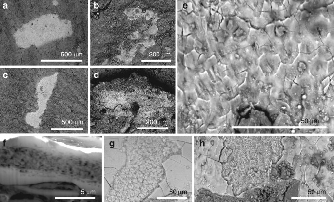
Small patches of tissue (0.01–0.4 mm2; Fig. 1a–d and Supplementary Figs. 26) are closely associated with fossil feathers (i.e., usually within 500 µm of carbonaceous feather residues, Supplementary Fig. 2e, g, j, k, o, s, t). The patches are definitively of fossil tissue, and do not reflect surface contamination with modern material during sample preparation, as they are preserved in calcium phosphate (see 'Taphonomy', below); further, several samples show margins that are overlapped, in part, by the surrounding matrix. The tissues have not, therefore, simply adhered to the sample surface as a result of contamination from airborne particles in the laboratory.
Fig. 1
Fig. 1
Phosphatised soft tissues in non-avian maniraptoran dinosaurs and a basal bird. ah Backscatter electron images of tissue in Confuciusornis (IVPP V 13171; a, e, f), Beipiaosaurus (IVPP V STM31-1; b, g), Sinornithosaurus (IVPP V 12811; c, h) and Microraptor (IVPP V 17972A; d). ad Small irregularly shaped patches of tissue. e Detail of tissue surface showing polygonal texture. f Focused ion beam-milled vertical section through the soft tissue showing internal fibrous layer separating two structureless layers. g, h Fractured oblique section through the soft tissues, showing the layers visible in f
The tissue patches are typically 3–6 µm thick and planar (Fig. 1a–e). Transverse sections and fractured surfaces show an inner fibrous layer (1.0–1.2 µm thick) between two thinner structureless layers (0.2–0.5 µm thick) (Fig. 1f–h). The external surface of the structureless layer is smooth and can show a subtle polygonal texture defined by polygons 10–15 µm wide (Fig. 1e, h).
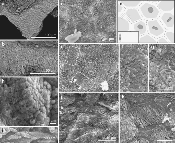
The fibrous layer also shows polygons (Figs. 1f, h and 2a–e, and Supplementary Fig. 6) that contain arrays of densely packed fibres 0.1–0.5 µm wide (Fig. 2f–i and Supplementary Fig. 5f). Well-preserved fibres show helicoidal twisting (Fig. 2h, i). Fibres in marginal parts of each polygon are 0.1–0.3 µm wide and oriented parallel to the tissue surface; those in the interior of each polygon are 0.3–0.5 µm wide and are usually perpendicular to the tissue surface (Fig. 2b, h and Supplementary Fig. S6d). In the marginal 1–2 µm of each polygon, the fibres are usually orthogonal to the lateral polygon margin and terminate at, or bridge the junction between, adjacent polygons (Fig. 2f, g and Supplementary Fig. 6e). The polygons are usually equidimensional but are locally elongated and mutually aligned, where the thick fibres in each polygon are sub-parallel to the tissue surface and the thin fibres, parallel to the polygon margin (Fig. 2j, k and Supplementary Fig. 6g–l). Some polygons show a central depression (Fig. 2c–e and Supplementary Fig. 6a–c) in which the thick fibres can envelop a globular structure 1–2 µm wide (Fig. 2e).
Fig. 2
Fig. 2
Ultrastructure of the soft tissues in Confuciusornis (IVPP V 13171). a, b Backscatter electron micrographs; all other images are secondary electron micrographs. a, b Closely packed polygons. c Detail of polygons showing fibrous contents, with d interpretative drawing. eg Polygon (e) with detail of regions indicated showing tonofibrils bridging (f) and abutting at (g) junction between polygons. h, i Helical coiling in tonofibrils. h Oblique view of polygon with central tonofibrils orientated perpendicular to the polygon surface. j, k Polygons showing stretching-like deformation

Fossil corneocytes

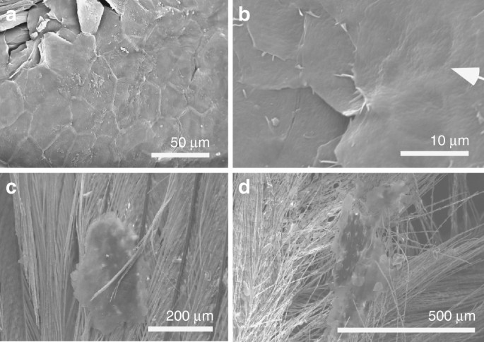
The texture of these fossil tissues differs from that of conchostracan shells and fish scales from the host sediment, the shell of modern Mytilus, modern and fossil feather rachis and modern reptile epidermis (Supplementary Fig. 7a–n). The elongate geometry of some polygons (Fig. 2j, k and Supplementary Fig. 6g, l) implies elastic deformation of a non-biomineralized tissue due to mechanical stress. On the basis of their size, geometry and internal structure, the polygonal structures are interpreted as corneocytes (epidermal keratinocytes). In modern amniotes, these are polyhedral-flattened cells (1–3 µm × ca. 15 µm) filled with keratin tonofibrils, lipids and matrix proteins18,19,20 (Fig. 3a, b and Supplementary Figs 2u–x, 8, 9). The outer structureless layer of the fossil material corresponds to the cell margin; it is thicker than the original biological template, i.e., the corneous cell envelope and/or cell membrane, but this is not unexpected, reflecting diagenetic overgrowth by calcium phosphate (see 'Taphonomy'). The fibres in the fossil corneocytes are identified as mineralised tonofibrils: straight, unbranching bundles of supercoiled α-keratin fibrils 0.25–1 µm wide18,21 that are the main component of the corneocyte cytoskeleton22 and are enveloped by amorphous cytoskeletal proteins22. In the fossils, the thin tonofibrils often abut those of the adjacent cell (Fig. 2g and Supplementary Fig. 6e), but locally can bridge the boundary between adjacent cells (Fig. 2f). The latter recalls desmosomes, regions of strong intercellular attachment between modern corneocytes23. The central globular structures within the fossil corneocytes resemble dead cell nuclei24, as in corneocytes of extant birds (but not extant reptiles and mammals)24 (Supplementary Fig. 8). The position of these pycnotic nuclei is often indicated by depressions in the corneocyte surface in extant birds24 (Fig. 3b); some fossil cells show similar depressions (Fig. 2c and Supplementary Fig. 6a–c).
Fig. 3
Fig. 3
Corneocytes in extant birds. ad Scanning electron micrographs of shed skin in extant zebra finch (Taeniopygia guttata (n = 1); ad). a Corneocytes defining polygonal texture. b Central depression (arrow) marks position of pycnotic nucleus. c, d Shed skin flakes entrained within feathers

Taphonomy

Keratin is a relatively recalcitrant biomolecule due to its heavily cross-linked paracrystalline structure and hydrophobic nonpolar character23. Replication of the fossil corneocytes in calcium phosphate is thus somewhat unexpected as this process usually requires steep geochemical gradients characteristic of early decay25 and usually applies to decay-prone tissues, such as muscle26 and digestive tissues27. Recalcitrant tissues such as dermal collagen can, however, be replicated in calcium phosphate where they contain an inherent source of calcium and, in particular, phosphate ions that are liberated during decay28. Corneocytes contain sources of both of these ions. During terminal differentiation, intracellular concentrations of calcium increase29 and α-keratin chains are extensively phosphorylated23. Further, corneocyte lipid granules30 are rich in phosphorus and phosphate31. These chemical moieties would be released during degradation of the granules and would precipitate on the remaining organic substrate, i.e., the tonofibrils.
In extant mammals, densely packed arrays of tonofibrils require abundant interkeratin matrix proteins for stability32. These proteins, however, are not evident in the fossils. This is not unexpected, as the proteins are rare in extant avian corneocytes33 and, critically, occur as dispersed monomers34 and would have a lower preservation potential than the highly cross-linked and polymerised keratin bundles of the tonofibrils. The outer structureless layer of the fossil corneocytes is thicker than the likely biological template(s), i.e., the corneous cell envelope (a layer of lipids, keratin and other proteins up to 100 nm thick that replaces the cell membrane during terminal differentiation34) and/or cell membrane. This may reflect a local microenvironment conducive to precipitation of calcium phosphate: during terminal differentiation, granules of keratohyalin, an extensively phosphorylated protein35 with a high affinity for calcium ions36, accumulate at the periphery of the developing corneocytes37. The thickness of the outer solid layer of calcium phosphate in the fossils, plus the gradual transition from this to the inner fibrous layer, suggests that precipitation of phosphate proceeded from the margins towards the interior of the corneocytes. In this scenario, phosphate availability in the marginal zones of the cells would have exceeded that required to replicate the tonofibrils. The additional phosphate would have precipitated as calcium phosphate in the interstitial spaces between the tonofibrils, progressing inwards from the inner face of the cell margin.

Skin shedding in feathered dinosaurs and early birds

In extant amniotes, the epidermal cornified layer is typically 5–20 cells thick (but thickness varies among species and location on the body38). The patches of fossil corneocytes, however, are one cell thick (Fig. 1f and Supplementary Figs. 5c, 10). This, plus the consistent small size (<400 a="" and="" continuous="" fidelity="" high="" i="" in="" inconsistent="" is="" m="" minority="" nbsp="" of="" patches="" preservation="" remarkably="" selective="" sheet="" situ="" the="" tissue.="" with="">n
= 8) of examples, the skin occurs at the edge of the sample of fossil soft tissues and thus could potentially represent a smaller fragment of an originally larger piece of fossil skin (with the remainder of the piece on the fossil slab). In most examples, however, the entire outline of the skin fragment is contained within the margin of a sample. Examination of the margins of various samples at high magnification reveals that the sample and surrounding sediment are often in exactly the same plane (e.g., Supplementary Fig. 10). Even where the margin of the sample of skin is covered by sediment, the sample is unlikely to have been much bigger than the apparent size as the fossil skin, being almost perfectly planar, forms a natural plane of splitting.There is no evidence that the preserved thickness of the skin is an artefact of preparation or erosion. During splitting of a rock slab, the plane of splitting frequently passes through the soft tissues in an uneven manner, exposing structures at different depths. In the fossils studied here, the plane of splitting usually passes through the corneocytes (exposing their internal structure), and rarely along the outer face of the corneocyte layer. There is no evidence for removal of more than one layer of corneocytes: FIB sections show preservation of only one layer and several SEM images show complete vertical sections through the preserved skin (where the relationship with the over- and underlying sediment is visible), with evidence for only a single layer of corneocytes. The fibrous internal fill of the fossil corneocytes is exposed where the plane of splitting of the fossil slab passes through the patches of tissue. The topography of the fossil corneocytes, however, varies with the position of the plane of splitting, which can vary locally through the soft tissues on a millimetre scale: the corneocytes can present with raised margins and a central depression, or with depressed margins and a central elevated zone (Fig. S9).
The size, irregular geometry and thickness of the patches of corneocytes resemble shed flakes of the cornified layer (dandruff-like particles39; Fig. 3). In extant birds, corneocytes are shed individually or in patches up to 0.5 mm2 that can be entrained within feathers (Fig. 3c, d and Supplementary Fig. 2u, v). The fossils described herein provide the first evidence for the skin shedding process in basal birds and non-avian maniraptoran dinosaurs and confirm that at least some non-avian dinosaurs shed their skin in small patches40. This shedding style is identical to that of modern birds18 (Fig. 3c, d) and mammals20 and implies continuous somatic growth. This contrasts with many extant reptiles, e.g., lepidosaurs, which shed their skin whole or in large sections21, but shedding style can be influenced by factors such as diet and environment41.

Evolutionary implications of fossil corneocyte structure

The fossil corneocytes exhibit key adaptations found in their counterparts in extant birds and mammals, especially their flattened polygonal geometry and fibrous cell contents consistent with α-keratin tonofibrils16. Further, the fossil tonofibrils (as in extant examples22) show robust intercellular connections and form a continuous scaffold across the corneocyte sheet (Fig. 2b, c, j and Supplementary Fig. 6). In contrast, corneocytes in extant reptiles contain a homogenous mass of β-keratin (with additional proteins present in the cell envelope) and fuse during development, forming mature β-layers without distinct cell boundaries42. The retention of pycnotic nuclei in the fossil corneocytes is a distinctly avian feature not seen in modern reptiles (but see ref. 20).
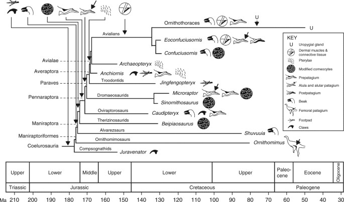
Epidermal morphogenesis and differentiation are considered to have diverged in therapsids and sauropsids31. Our data support other evidence that shared epidermal features in birds and mammals indicate convergent evolution43 and suggest that lipid-rich corneocyte contents may be evolutionarily derived characters in birds and feathered non-avian maniraptorans. Evo-devo studies have suggested that the avian epidermis could have arisen from the expansion of hinge regions in ‘protofeather’-bearing scaly skin20. While fossil evidence for this transition is lacking, our data show that the epidermis of basal birds and non-avian maniraptoran dinosaurs had already evolved a decidedly modern character, even in taxa not capable of powered flight. This does not exclude the possibility that at least some of the epidermal features described here originated in more basal theropods, especially where preserved skin lacks evidence of scales (as in Sciurumimus44). Refined genomic mechanisms for modulating the complex expression of keratin in the epidermis45, terminal differentiation of keratinocytes and the partitioning of α- and β-keratin synthesis in the skin of feathered animals32 were probably modified in tandem with feather evolution close to the base of the Maniraptora by the late Middle Jurassic (Fig. 4). Existing fossil data suggest that this occurred after evolution of the beak in Maniraptoriformes and before evolution of the forelimb patagia and pterylae (Fig. 4); the first fossil occurrences of all of these features span ca. 10–15 Ma, suggesting a burst of innovation in the evolution of feathered integument close to and across the Lower-Middle Jurassic boundary. The earliest evidence for dermal musculature associated with feathers is ca. 30 Ma younger, in a 125 Ma ornithothoracean bird17. Given the essential role played by this dermal network in feather support and control of feather orientation18, its absence in feathered non-avian maniraptorans may reflect a taphonomic bias.
Fig. 4
Fig. 4
Schematic phylogeny, scaled to geological time, of selected coelurosaurs showing the pattern of acquisition of key modifications of the skin. The phylogeny is the most likely of the maximum likelihood models, based on minimum-branch lengths (mbl) and transitions occurring as all-rates-different (ARD). Claws and footpads are considered primitive in coelurosaurs. Available data indicate that modified keratinocytes, and continuous shedding, originated close to the base of the Maniraptora; this is predicted to shift based on future fossil discoveries towards the base of the Coelurosauria to include other feathered taxa
In certain aspects, the fossil corneocytes are distinctly non-avian and indicate that feathered dinosaurs and early birds had a unique integumentary anatomy and physiology transitional between that of modern birds and non-feathered dinosaurs. In extant birds, corneocyte tonofibrils are dispersed loosely among intracellular lipids19; this facilitates evaporative cooling in response to heat production during flight and insulation by plumage46. In contrast, the fossil tonofibrils are densely packed and fill the cell interior. There is no evidence for post-mortem shrinkage of the fossil corneocytes: the size range is consistent with those in modern birds, and there is no evidence for diagenetic wrinkling, contortion or separation of individual cells. This strongly suggests that the preserved density of tonofilaments in the fossil corneocytes reflects originally higher densities than in extant birds. This is not a function of body size: extant birds of disparate size (e.g., zebra finch and ostrich) exhibit loosely dispersed tonofibrils47. The fossil birds are thus likely to have had a lower physiological requirement for evaporative cooling and, in turn, lower body heat production related to flight activity46 than in modern birds. This is consistent with other evidence for low basal metabolic rates in non-avian maniraptoran dinosaurs47,48 and basal birds47 and with hypotheses that the feathers of Microraptor49 and, potentially, Confuciusornis48 (but see ref. 50) were not adapted for powered flight, at least for extended periods50.

Methods

Fossil material

This study used the following specimens in the collections of the Institute for Vertebrate Palaeontology and Paleanthropology, Beijing, China: Confuciusornis (IVPP V 13171), Beipiaosaurus (IVPP V STM31-1), Sinornithosaurus (IVPP V 12811) and Microraptor (IVPP V 17972A). Small (2–10 mm2) chips of soft tissue were removed from densely feathered regions of the body during initial preparation of the specimens and stored for later analysis. Precise sampling locations are not known.

Modern bird tissues

Male specimens of the zebra finch Taeniopygia guttata (n = 1) and the Java sparrow Lonchura oryzivora (n = 2) were euthanased via cervical dislocation. Individual feathers dissected from T. guttata and moulted down feathers from a male specimen of the American Pekin duck (Anas platyrhynchos domestica) were not treated further. Small (ca. 10–15 mm2) pieces of skin and underlying muscle tissue were dissected from the pterylae of the breast of reproductively active male specimens of L. oryzivora raised predominantly on a diet of seeds in October 2016. Tissue samples were fixed for 6 h at 4 °C in 4% paraformaldehyde. After snap freezing in isopentane, tissue was coronal sectioned (10 µm thickness) with a Leica CM1900 cryostat. All sections were allowed to air dry at room temperature for 3 h and stored at −80 °C prior to immunohistology.

Ethics

The authors have complied with all relevant ethical regulations. Euthanasia of T. guttata and L. oryzivora was approved by the Health Products Regulatory Authority of Ireland via authorisation AE19130-IO87 (for T. guttata) and CRN 7023925 (for L. oryzivora).

Electron microscopy

Samples of soft tissues were removed from fossil specimens with sterile tools, placed on carbon tape on aluminium stubs, sputter coated with C or Au and examined using a Hitachi S3500-N and a FEI Quanta 650 FEG SEM at accelerating voltages of 5–20 kV.
Untreated feathers and fixed and dehydrated samples of skin from modern birds were placed on carbon tape on aluminium stubs, sputter coated with C or Au and examined using a Hitachi S3500-N and a FEI Quanta 650 FEG SEM at accelerating voltages of 5–20 kV. Selected histological sections of L. oryzivora were deparaffinized in xylene vapour for 3 × 5 min, sputter coated with Au, and examined using a FEI Quanta 650 FEG SEM at an accelerating voltage of 15 kV. The brightness and contrast of some digital images were adjusted using Deneba Canvas software.

Focussed ion beam-scanning electron microscopy

Selected samples of fossil tissue were analysed using an FEI Quanta 200 3D FIB-SEM. Regions of interest were coated with Pt using an in situ gas injection system and then milled using Ga ions at an accelerating voltage of 30 kV and a beam current of 20 nA–500 pA.

Immunohistology

Histological sections were incubated in permeabilization solution (0.2% Triton X-100 in 10 mM phosphate-buffered saline (PBS)) for 30 min at room temperature, washed once in 10 mM PBS and blocked in 5% normal goat serum in 10 mM PBS for 1 h at room temperature. Sections were incubated in primary antibody to cytokeratin (1:300; ThermoFisher) in 2% normal goat serum in 10 mM PBS overnight at 4 °C. Following. 3 × 5 min wash in 10 mM PBS, sections were incubated with a green fluorophore-labelled secondary antibody (1:500; Invitrogen) for 2 h at room temperature. After a 3 × 10 min wash in 10 mM PBS, sections were incubated in BisBenzimide nuclear counterstain (1:3000; Sigma-Aldrich) for 4 min at room temperature. Sections were washed briefly, mounted and coverslipped with PVA-DABCO.

Confocal microscopy

Digital images were obtained using an Olympus AX70 Provis upright fluorescence microscope and a ×100 objective and stacked using Helicon Focus software (www.heliconfocus.com).

Data availability

The data that support the findings of this study can be downloaded from the CORA repository (www.cora.ucc) at http://hdl.handle.net/10468/5795.

Additional information

Publisher's note: Springer Nature remains neutral with regard to jurisdictional claims in published maps and institutional affiliations.

References

  1. 1.
    Sawyer, R. H., Knapp, L. W. & O’Guin, W. M. in Biology of the Integument 2: Vertebrates (eds Bereiter-Hahn, J. B. & Matoltsy, A. G.) 194–238 (Springer, Heidelberg, 1986).
  • 2.
    Landmann, L. in Biology of the Integument 2: Vertebrates (eds Bereiter-Hahn, J. B. & Matoltsy, A. G.) 153–187 (Springer, New York, 1986).
  • 3.
    Menon, G. K., Bown, B. E. & Elias, P. M. Avian epidermal differentiation: role of lipids in permeability barrier formation. Tissue Cell 18, 71–82 (1986).
  • 4.
    Prum, R. O. & Brush, A. H. The evolutionary origin and diversification of feathers. Quart. Rev. Biol. 77, 261–295 (2002).
  • 5.
    Xu, X., Zheng, F. & You, H. Exceptional dinosaur fossils show ontogenetic development of early feathers. Nature 464, 1338–1341 (2010).
  • 6.
    Norell, M. A. & Xu, X. Feathered dinosaurs. Annu. Rev. Earth Planet. Sci. 33, 277–299 (2005).
  • 7.
    Godefroit, P. et al.. Jurassic ornithischian dinosaur from Siberia with both feathers and scales. Science 345, 451–455 (2014).
  • 8.
    Alibardi, L. & Gill, B. J. Epidermal differentiation in embryos of the tuatara Sphenodon punctatus (Reptilia, Sphenodontidae) in comparison with the epidermis of other reptiles. J. Anat. 211, 92–103 (2007).
  • 9.
    Wu, P. et al. Evo-devo of amniote integuments and appendages. Int. J. Dev. Biol. 48, 249–270 (2004).
  • 10.
    Martill, D. M., Batten, D. J. & Loydell, D. K.. new specimen of the dinosaur cf. Scelidosaurus with soft tissue preservation. Palaeontology 43, 549–559 (2000).
  • 11.
    Lingham-Soliar, T. & Plodowski, G. in The integument of Psittacosaurus from Liaoning Province, China: taphonomy, epidermal patterns and color of. ceratopsian dinosaur. Naturwissenschaften 97, 479–486 (2010).
  • 12.
    Martill, D. M. Organically preserved dinosaur skin: taphonomic and biological implications. Mod. Geol. 16, 61–68 (1991).
  • 13.
    Manning, P. L. et al. Mineralized soft-tissue structure and chemistry in. mummified hadrosaur from the Hell Creek Formation, North Dakota (USA). Proc. Biol. Sci. 276, 3429–3437 (2009).
  • 14.
    Schweitzer, M. H. Soft tissue preservation in terrestrial Mesozoic vertebrates. Annu. Rev. Earth Planet. Sci. 39, 187–216 (2011).
  • 15.
    Chiappe, L. M. et al. Sauropod dinosaur embryos from the Late Cretaceous of Patagonia. Nature 396, 258–260 (1998).
  • 16.
    McNamara, M. E. et al. Reconstructing carotenoid-based and structural coloration in fossil skin. Curr. Biol. 26, 1075–1082 (2016).
  • 17.
    Navalón, G., Marugán-Lobón, J., Chiappe, L. M., Sanz, J. L. & Buscalioni, A. D. Soft-tissue and dermal arrangement in the wing of an Early Cretaceous bird: implications for the evolution of avian flight. Sci. Rep. 5, 14864 (2015).
  • 18.
    Lucas, A. M. & Stettenheim, P. R. Avian Anatomy: Integument (U.S. Department of Agriculture, Washington, 1972).
  • 19.
    Menon, G. K. & Menon, J. Avian epidermal lipids: functional considerations and relationship to feathering. Am. Zool. 40, 540–552 (2000).
  • 20.
    Alibardi, L. Adaptation to the land: the skin of reptiles in comparison to that of amphibians and endotherm amniotes. J. Exp. Zool. 298B, 12–41 (2003).
  • 21.
    Ramms, L. Keratins as the main component for the mechanical integrity of keratinocytes. Proc. Natl Acad. Sci. USA 110, 18513–18518 (2013).
  • 22.
    Ishida, A. et al. Sequential reorganization of cornifed cell keratin filaments involving filaggrin-mediated compaction and keratin deimination. J. Invest. Dermatol. 118, 282–287 (2002).
  • 23.
    Skerrow, D. in Biology of the Integument 2: Vertebrates (eds Bereiter-Hahn, J. B. & Matoltsy, A. G.) (Springer, Heidelberg, 1986).
  • 24.
    Mihara, M. Scanning electron microscopy of skin surface and the internal structure of corneocyte in normal human skin. An application of the osmium-dimethyl sulfoxide-osmium method. Arch. Dermatol. Res. 280, 293–299 (1988).
  • 25.
    Sagemann, J., Bale, D. J., Briggs, D. E. G. & Parkes, R. J. Controls on the formation of authigenic minerals in association with decaying organic matter: an experimental approach. Geochem. Cosmochim. Acta 68, 1083–1095 (1999).
  • 26.
    Wilby, P. R. & Briggs, D. E. G. Taxonomic trends in the resolution of detail preserved in fossil phosphatized soft tissues. Geobios 20, 493–502 (1997).
  • 27.
    Butterfield, N. J. Leanchoilia guts and the interpretation of three-dimensional structures in Burgess Shale-type fossils. Paleobiology 28, 155–171 (2002).
  • 28.
    McNamara, M. E. et al. Soft-tissue preservation in Miocene frogs from Libros, Spain: insights into the genesis of decay microenvironments. Palaios 24, 104–117 (2009).
  • 29.
    Farage, M. A., Miller, K. W. & Maibach, H. I. Textbook of Aging Skin (Heidelberg, Springer, 2010).
  • 30.
    Alibardi, L. Immunocytochemical and autoradiographic studies on the process of keratinization in avian epidermis suggests absence of keratohyalin. J. Morphol. 259, 238–253 (2004).
  • 31.
    Alibardi, L. & Toni, M. Cytochemical, biochemical and molecular aspects of the process of keratinization in the epidermis of reptilian scales. Prog. Histochem. Cytochem. 40, 73–134 (2006).
  • 32.
    Fukuyama, K. & Epstein, W. L. Protein synthesis studied by autoradiography in the epidermis of different species. Am. J. Anat. 122, 269–273 (1968).
  • 33.
    Candi, E., Schmidt, R. & Melino, G. The cornified envelope:. model of cell death in the skin. Nat. Rev. Mol. Cell Biol. 6, 328–340 (2005).
  • 34.
    van der Reest, A. J., Wolfe, A. P. & Currie, P. J. Reply to comment on: “a densely feathered ornithomimid (Dinosauria: Theropoda) from the Upper Cretaceous Dinosaur Park Formation, Alberta, Canada. Cret. Res. 58, 108–117 (2016).
  • 35.
    Parakkal, P. F. & Alexander, N. J. Keratinization:. Survey of Vertebrate Epithelia. (Academic Press, New York, 2012).
  • 36.
    Markova, N. G. et al. Profilaggrin is. major epidermal calcium-binding protein. Mol. Cell. Biol. 13, 613–625 (1993).
  • 37.
    Schachner, L. A. & Hansen, R. C. Pediatric Dermatology (Elsevier, Mosby, 2011).
  • 38.
    Eurell, J. A. & Frappier, B. L. Textbook of Veterinary Histology (Blackwell, Oxford, 2013).
  • 39.
    Stettenheim, P. in Avian Biology (eds Farner, D. S. & King, J. R.) 2–63 (Acadmic Press, New York, 1972).
  • 40.
    Poinar, G. P. Jr & Poinar, R. What Bugged the Dinosaurs? Insects, Disease and Death in the Cretaceous (Princeton University Press, Princeton, 2008).
  • 41.
    Shankar, P. G. & Whitaker, N. Ecdysis in the king cobra. Russ. J. Herpet. 16, 1–5 (2009).
  • 42.
    Alibardi, L. & Toni, M. Characterization of keratins and associated proteins involved in the corneification of crocodilian epidermis. Tissues Cell 39, 311–323 (2007).
  • 43.
    Maderson, P. F. A. & Alibardi, L. The development of the sauropsid integument:. contribution to the problem of the origin and evolution of feathers. Am. Zool. 40, 513–529 (2000).
  • 44.
    Rauhut, O. W. M., Foth, C., Tischlinger, H. & Norell, M. A. Exceptionally preserved juvenile megalosauroid theropod dinosaur with filamentous integument from the Late Jurassic of Germany. Proc. Natl Acad. Sci. USA 109, 11746–11751 (2012).
  • 45.
    Porter, R. M. & Lane, E. B. Phenotypes, genotypes and their contribution to understanding keratin function. Trends Genet. 19, 278–285 (2003).
  • 46.
    Menon, G. K. et al. Ultrastructural organization of avian stratum corneum lipids as the basis for facultative cutaneous waterproofing. J. Morphol. 227, 1–13 (1996).
  • 47.
    Xu, X. et al. An integrative approach to understanding bird origins. Science 346, 153293–153210 (2014).
  • 48.
    Nudds, R. L. & Dyke, G. J. Narrow primary feather rachises in Confuciusornis and Archaeopteryx suggest poor flight ability. Science 328, 887–889 (2010).
  • 49.
    Dyke, G. J., de Kat, R., Palmer, C., van der Kindere, J. & Naish, B. Aerodynamic performance of the feathered dinosaur Microraptor and the evolution of feathered flight. Nat. Commun. 4, 2489 (2013).
  • 50.
    Falk, A. R., Kaye, T. G., Zhou, Z. & Burnham, D. A. Laser fluorescence illuminates the soft tissue and life habits of the Early Cretaceous bird Confuciusornis. PLoS ONE 11, e0167284 (2016).
  • Acknowledgements

    We thank Zheng Fang, Laura and Jonathan Kaye, Vince Lodge, Giliane Odin and Luke Harman for assistance. Funded by a European Research Council Starting Grant (ERC-2014-StG-637691-ANICOLEVO) and Marie Curie Career Integration Grant (FP7-2012-CIG-618598) to M.E.M., Natural Environment Research Council Grants (NE/E011055/1 and 1027630/1) to M.J.B. and S.K. and by grants from the Royal Society (H.C. Wong Postdoctoral Fellowship), National Natural Sciences Foundation of China (41125008 and 41688103), Chinese Academy of Sciences (KZCX2-EW-105) and Linyi University to F.Z. and X.X.

    Author information

    Affiliations

    1. School of Biological, Earth and Environmental Sciences, University College Cork, North Mall, Cork, T23 TK30, Ireland

      • Maria E. McNamara
      •  & Chris S. Rogers
    2. Institute of Geology and Paleontology, Linyi University, Shuangling Road, Linyi City, Shandong, 276005, China

      • Fucheng Zhang
    3. School of Earth Sciences, University of Bristol, Queen’s Road, Bristol, BS8 1RJ, UK

      • Stuart L. Kearns
      •  & Michael J. Benton
    4. UCD School of Earth Sciences, University College Dublin, Dublin, D04 N2E5, Ireland

      • Patrick J. Orr
    5. Department of Anatomy and Neuroscience, University College Cork, Western Road, Cork, T12 XF62, Ireland

      • André Toulouse
      •  & Tara Foley
    6. School of Biological and Chemical Sciences, Queen Mary University of London, Mile End Rd., London, E1 4NS, UK

      • David W. E. Hone
    7. School of Physical Sciences, The Open University, Walton Hall, Milton Keynes, MK7 6AA, UK

      • Diane Johnson
    8. Institute of Vertebrate Paleontology and Paleoanthropology, 142 Xizhimenwai St., 100044, Beijing, China

      • Xing Xu
      •  & Zhonghe Zhou

    Contributions

    M.E.M., F.Z., P.J.O., S.L.K. and M.J.B. designed the study; M.E.M. and F.Z. carried out SEM analyses with assistance from P.J.O., S.L.K., D.W.E.H. and C.S.R.; D.J., P.J.O. and S.L.K. carried out FIB-SEM analyses; A.T., T.F. and M.E.M. carried out fluorescence microscopy; M.E.M., P.J.O., S.L.K. and M.J.B. wrote the paper with contributions from all authors.

    Competing interests

    The authors declare no competing interests.

    Corresponding authors

    Correspondence to Maria E. McNamara or Fucheng Zhang or Xing Xu.

    Electronic supplementary material

    About this article

    Publication history

    Received

    Accepted

    Published

    DOI