Giant
tortoises are among the longest-lived vertebrate animals and, as such,
provide an excellent model to study traits like longevity and
age-related diseases. However, genomic and molecular evolutionary
information on giant tortoises is scarce. Here, we describe a global
analysis of the genomes of Lonesome George—the iconic last member of Chelonoidis abingdonii—and the Aldabra giant tortoise (Aldabrachelys gigantea).
Comparison of these genomes with those of related species, using both
unsupervised and supervised analyses, led us to detect lineage-specific
variants affecting DNA repair genes, inflammatory mediators and genes
related to cancer development. Our study also hints at specific
evolutionary strategies linked to increased lifespan, and expands our
understanding of the genomic determinants of ageing. These new genome
sequences also provide important resources to help the efforts for
restoration of giant tortoise populations.
Main
Comparative
genomic analyses leverage the mechanisms of natural selection to find
genes and biochemical pathways related to complex traits and processes.
Multiple works have used these techniques with the genomes of long-lived
mammals to shed light on the signalling and metabolic networks that
might play a role in regulating age-related conditions1,2.
Similar studies on unrelated longevous organisms might unveil novel
evolutionary strategies and genetic determinants of ageing in different
environments. In this regard, giant tortoises constitute one of the few
groups of vertebrates with an exceptional longevity: in excess of
100 years according to some estimates.
In this manuscript, we
report the genomic sequencing and comparative genomic analysis of two
long-lived giant tortoises: Lonesome George—the last representative of Chelonoidis abingdonii3, endemic to the island of Pinta (Galapagos Islands, Ecuador)—and an individual of Aldabrachelys gigantea, endemic to the Aldabra Atoll and the only extant species of giant tortoises in the Indian Ocean4 (Fig. 1a).
Unsupervised and supervised comparative analyses of these genomic
sequences add new genetic information on the evolution of turtles, and
provide novel candidate genes that might underlie the extraordinary
characteristics of giant tortoises, including their gigantism and
longevity.
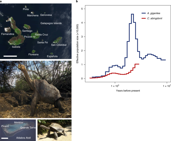
Fig. 1: Geographical and temporal distribution of giant tortoises.
a,
Satellite view of the Galapagos Islands (top; scale bar: 50 km) and
Aldabra Atoll (bottom left; scale bar: 10 km), and pictures of C. abingdonii (middle) and A. gigantea (bottom right). Both pictures are from http://eol.jsc.nasa.gov. b,
Demographic history of giant tortoises, inferred using a hidden Markov
model approach as implemented in the PSMC model. The default mutation
rate (μ) for humans of 2.5 × 10⁻⁸ and an average generation time (g) of 25 years were used in the calculations.
The genome of Lonesome George was sequenced using a combination of Illumina and PacBio platforms (Supplementary Section 1.1).
The assembled genome (CheloAbing 1.0) has a genomic size of
2.3 gigabases and contains 10,623 scaffolds with an N50 of
1.27 megabases (Supplementary Section 1.1 and Supplementary Tables 1–3). We also sequenced, with the Illumina platform, the closely related tortoise A. gigantea at an average read depth of 28×. These genomic sequences were aligned to CheloAbing 1.0.
TimeTree database estimations (http://www.timetree.org)
indicate that Galapagos and Aldabra giant tortoises shared a last
common ancestor about 40 million years ago, while both diverged from the
human lineage more than 300 million years ago (Supplementary Section 1.4). A preliminary analysis of demographic history using the pairwise sequentially Markovian coalescent (PSMC)5 model showed that while the effective population size of C. abingdonii
has been steadily declining for the past million years, with a slight
uptick about 90,000 years ago, the population of Aldabra giant tortoises
experienced substantial fluctuations over this period (Fig. 1b). Effective population size reconstructions for C. abingdonii
lose statistical power at the million-year time frame, probably due to
complete coalescence. In turn, this suggests that overall diversity in
these giant tortoises must have been low throughout many generations.
Together, these results prompt us to propose that the populations of
these insular giant tortoises were vulnerable at the time of human
discovery of the Galapagos Islands, probably elevating their extinction
risk.
Using homology searches with known gene sets from humans and Pelodiscus sinensis (the Chinese soft-shell turtle), along with RNA sequencing (RNA-Seq) data from C. abingdonii blood and an A. gigantea granuloma, we automatically predicted a primary set of 27,208 genes from the genome assembly using the MAKER2 algorithm6.
We then performed pairwise alignments between each of the primary
predicted protein sequences and the UniProt databases for humans and P. sinensis, whose annotated sequences show relatively high quality when compared with data available for other turtles7.
Using alignments spanning at least 80% of the longest protein and
showing more than 60% identity, we constructed sets of protein families
shared among these species. This preliminary analysis singled out
several protein families that seem to have undergone moderate expansion
in a common ancestor of C. abingdonii and A. gigantea. Almost all of these expansions were also confirmed in the genome of the related, long-lived tortoise Gopherus agassizii (Supplementary Section 1.2 and Supplementary Table 4).
Most of these genes have been linked to exosome formation, suggesting
that this process may have been important in tortoise evolution.
We
also interrogated the predicted gene set for evidence of positive
selection in giant tortoises. This analysis singled out 43 genes with
evidence of giant-tortoise-specific positive selection (Supplementary
Section 1.2, Supplementary Table 5 and Supplementary Fig. 1). This list includes genes with known roles in the dynamics of the tubulin cytoskeleton (TUBE1 and TUBG1) and intracellular vesicle trafficking (VPS35). Importantly, the analysis of genes showing evidence of positive selection also includes AHSG and FGF19, whose expression levels have been linked to successful ageing in humans8. The role of both factors in metabolism regulation9,10—another hallmark of ageing11,12—suggests
that the specific changes observed in these proteins may have arisen to
accommodate the challenges that longevity poses on this system. The
list of genes with signatures of positive selection also features TDO2,
whose inhibition has been proposed to protect against age-related
diseases through regulation of tryptophan-mediated proteostasis13. In addition, we found evidence for positive selection affecting several genes involved in immune system modulation, such as MVK, IRAK1BP1 and IL1R2.
Taken together, these results identify proteostasis, metabolism
regulation and immune response as key processes during the evolution of
giant tortoises via effects on longevity and resistance to infection.
Parallel
to this automatic analysis, we used manually supervised annotation on
more than 3,000 genes selected a priori for a series of
hypothesis-driven studies on development, physiology, immunity,
metabolism, stress response, cancer susceptibility and longevity
(Supplementary Section 1.3 and Supplementary Fig. 2).
We searched for truncating variants, variants affecting known motifs
and variants whose human counterparts are related to known genetic
diseases (Supplementary Section 1.3 and Supplementary Table 6).
These variants were first confirmed with the RNA-Seq data. Then, more
than 100 of the most interesting variants in terms of putative
functional relevance were also validated by PCR amplification followed
by Sanger sequencing. To this end, we used a panel of genomic DNA
samples of 11 different species of giant tortoises endemic to different
islands from the Galapagos Archipelago (Supplementary Section 1, Supplementary Table 7 and Supplementary Fig. 3).
The
manually supervised annotation of development-related genes showed the
complete conservation of the Hox gene set among giant tortoises, with
the exception of HOXC3, which seems to have been lost in the radiation of Archelosauria14,15 (Supplementary Section 2, Supplementary Table 8 and Supplementary Fig. 4). BMP and GDF gene families were also found to be conserved, although the duplication event that gave rise to GDF1 and GDF3 in mammals did not occur in turtles, birds and crocodiles. In contrast, we found a duplication of the ParaHox gene CDX4
in giant tortoises, also present in other reptiles as well as avian
reptiles (birds). This annotation also showed the duplication of WNT11 in turtles and chickens (but not in the lizard Anolis carolinensis), and the specific duplication of WNT4
in turtles. Given the roles of these duplicated genes and their
conservation in most vertebrate species, they could prove to be useful
candidates to study the morphological development of turtles,
particularly in relation to shell formation. Of note, KDSR—one of the genes possibly under positive selection in giant tortoises—has been linked to hyperkeratinization disorders16. Also, in this regard, we annotated 30 β-keratins in C. abingdonii, 26 of which seem to be functional. These numbers are lower than those previously reported for β-keratins in other turtles17. Finally, we did not find in C. abingdonii or A. gigantea any functional orthologues of genes specifically involved in tooth development (such as ENAM, AMEL, AMBN, DSPP, KLK4 and MMP20).
This finding confirms a pattern in the evolutionary molecular
mechanisms for tooth loss, which seems to have been followed
consistently and independently across vertebrates. Taken together, these
results offer multiple candidates to study developmental traits in
tortoises (Supplementary Section 2 and Supplementary Figs. 5–8).
In
most species, the immune function is an evolutionary driver that is
under strong selective pressure and has important implications in ageing
and disease18.
The specific components and functionality of immune system components
in Reptilia, however, have not been extensively characterized beyond the
major histocompatibility complex (MHC)19,20.
Our detailed analysis of 891 genes involved in immune function
consistently found duplications affecting immunity genes in giant
tortoises compared with mammals (Supplementary Section 3, Supplementary Table 9 and Supplementary Figs. 9–13). We found a genomic expansion of PRF1 (encoding perforin) in giant tortoises and other turtles, compared with chickens (one copy), A. carolinensis (two copies) and most mammals (one copy). Both C. abingdonii and A. gigantea possess 12 copies of this gene (validated by Sanger sequencing), although three of them have been pseudogenized in C. abingdonii.
In addition, we detected and validated, by Sanger sequencing, an
expansion of the chymase locus, containing granzymes, in giant tortoises
(Supplementary Section 3.1 and Supplementary Fig. 10).
Both expansions are expected to affect cytotoxic T lymphocyte and
natural killer functions, which play important roles in defence against
both pathogens and cancer21,22.
Other concurrent expansions involve APOBEC1, CAMP, CHIA and NLRP
genes, which participate in viral, microbial, fungal and parasite
defence, respectively. These results suggest that the innate immune
system in turtles, and especially in giant tortoises, may play a more
relevant role than in mammals, consistent with the less important role
that adaptive immunity seems to play19.
We found that class I and II MHC genes probably underwent a duplication
event in a common ancestor between giant tortoises and painted turtles (Chrysemys picta bellii).
We also annotated 40 class III MHC genes, thus confirming the
conservation of this cluster in giant tortoises. The large number of MHC
genes in giant tortoises is consistent with the suggestion that
ancestors of archosaurs and chelonians did not possess a minimal
essential MHC as found in the chicken genome20 (Supplementary Section 3.3, Supplementary Table 10 and Supplementary Figs. 14–16).
Giant tortoises are at the upper end of the size scale for extant Chelonii, and have often been used as an example of gigantism23. We analysed a series of genes involved in size regulation in vertebrates, most notably dogs (Supplementary Section 2, Supplementary Table 8 and Supplementary Fig. 6).
Our results on genes related to growth hormone, the insulin-like growth
factor (IGF) system and stanniocalcins suggest that these genes are
well conserved; therefore, additional size determinants may exist in
giant tortoises. As a complex phenotype, gigantism in tortoises is
expected to be caused by interactions between different genetic and
environmental factors. An interesting finding in this regard is the
presence of several gene variants in tortoises (including G. agassizii) probably affecting the activities of glucose metabolism genes, such as MIF (p.N111C; expected to yield a locked trimer) and GSK3A
(p.R272Q in the activation loop). Given the roles of these positions in
the mammalian orthologues of these genes, tortoise-specific changes
could point to differences in the regulation of glucose intake and
tolerance (Supplementary Section 4, Supplementary Table 11, and Supplementary Figs. 17 and 18).
We also found expansions and inactivations in other genes involved in
energy metabolism. Thus, glyceraldehyde-3-phosphate dehydrogenase (GAPDH)—a glycolytic enzyme with a key role in energy production, as well as in DNA repair and apoptosis24—is expanded in giant tortoises. Conversely, the NLN
gene encoding neurolysin is pseudogenized in tortoises. The loss of
this gene in mice has been related to improved glucose uptake and
insulin sensitivity25.
Taken together, these results led us to hypothesize that genomic
variants affecting glucose metabolism may have been a factor in the
development of tortoises.
The analysis of genes related to the
stress response has also highlighted several putative variants in giant
tortoises affecting globins and DNA repair factors (Supplementary
Section 5, Supplementary Tables 12 and 13, and Supplementary Figs. 19–22, 32 and 33). We found that, despite living terrestrially, giant tortoises conserve the hypoxia-related globin GbX26.
Together with coelacanths, turtles, including giant tortoises, are the
only organisms known to possess all eight different types of globins27. Consistent with this, we found in both giant tortoise genomes a variant in the transcription factor TP53 (p.S106E) that has been linked to hypoxia resistance in some mammals and fishes28.
The presence of the same residue in Testudines strongly suggests a
process of convergent evolution in the adaptation to hypoxia, probably
driven by an ancestral aquatic environment, which left this footprint in
the genomes of terrestrial giant tortoises.
An important trait of
large, long-lived vertebrates is their need for tighter cancer
protection mechanisms, as illustrated by Peto’s paradox29,30.
In turn, this need for additional protection illustrates the deep
relationship and interdependence between cancer and longevity (Fig. 2). Notably, tumours are believed to be very rare in turtles31.
Therefore, we analysed more than 400 genes classified in a
well-established census of cancer genes as oncogenes and tumour
suppressors32.
Although most presented a highly conserved amino acid sequence when
compared with the sequences of other organisms, we uncovered alterations
in several tumourigenesis-related genes (Fig. 2a, Supplementary Section 6, Supplementary Table 14 and Supplementary Figs. 23–29).
First, we found that several putative tumour suppressors are expanded
in turtles compared with other vertebrates, including duplications in SMAD4, NF2, PML, PTPN11 and P2RY8. In addition, the aforementioned expansion of PRF1, together with the tortoise-specific duplication of PRDM1,
suggests that immunosurveillance may be enhanced in turtles. Likewise,
we found giant-tortoise-specific duplications affecting two putative
proto-oncogenes—MYCN and SET. Notably, the SET complex
mediates oxidative stress responses induced by mitochondrial damage
through the action of PRF1 and GZMA in cytotoxic T lymphocyte- and
natural killer-mediated cytotoxicity33.
Taken together, these results suggest that multiple gene copy-number
alterations may have influenced the mechanisms of spontaneous tumour
growth. Nevertheless, further studies are needed to evaluate the genomic
determinants of putative giant-tortoise-specific cancer mechanisms.
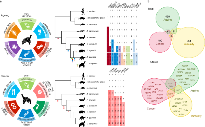
Fig. 2: Genomic basis of longevity and cancer in giant tortoises.
a, Genes potentially implicated in C. abingdonii and A. gigantea
longevity extension and cancer resistance, classified according to
their putative role in the different hallmarks. Tables indicate
copy-number variations and relevant variants of age-related genes and
tumour suppressors found in C. abingdonii, A. gigantea and
other species. Within these tables, numbers indicate gene copy numbers,
and asterisks represent pseudogenization events. Dots in colours
relating to each hallmark represent presence of the variant. b,
Venn diagrams showing the relationships between cancer-, ageing- and
immunity-related genes, as classified before annotation. Top, all of the
genes related to each category that have been manually annotated,
including the number of genes in each group. Bottom, those genes showing
potentially interesting variations after annotation.
Finally,
we selected, for manually supervised annotation, a set of 500 genes
that may be involved in ageing modulation (Supplementary Section 7 and Supplementary Table 15). The extreme longevity of giant tortoises is expected to involve multiple genes affecting different hallmarks of ageing11.
We found several alterations in the genomes of giant tortoises that may
play a direct role in six of them, and impinge on other ageing
hallmarks and processes, such as cancer progression34 (Fig. 2b).
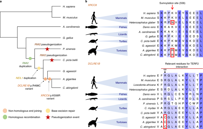
First, we identified changes in three candidate factors (NEIL1, RMI2
and XRCC6) related to the maintenance of genome integrity, a primary
hallmark of ageing11 (Fig. 3a).
Thus, we found and validated a duplication affecting NEIL1, a key
protein involved in the base-excision repair process whose expression
has been linked to extended lifespans in several species35. Likewise, RMI2
is duplicated in tortoises, suggesting an enhanced ability to resolve
homologous recombination intermediates to limit DNA crossover formation
in cells36.
In a preliminary exploration of this hypothesis, we overexpressed NEIL1 and RMI2 in HEK-293T cells and exposed the infected cells to a sublethal dosage of H2O2
or ultraviolet light, monitoring DNA damage by western blot analysis at
24 and 48 h after treatment. As shown in Supplementary Figs. 22, 32 and 33,
the expression of both genes results in reduced levels of
phosphorylated histone H2AX and cleaved poly (ADP-ribose) polymerase
(PARP), suggesting reduced levels of DNA damage37.
In turn, this result is consistent with the hypothesis that NEIL1 and
RMI2 levels may regulate the strength of DNA repair mechanisms. Also in
relation to DNA repair mechanisms, we identified and validated a variant
affecting XRCC6—encoding a helicase involved in non-homologous
end joining of double-strand DNA breaks—which may affect a known
sumoylation site (p.K556R). This lysine is conserved in diverse
vertebrates but, notably, is changed in giant tortoises, and also in the
naked mole rat (p.K556N), the longest-lived rodent, which suggests a
putative process of convergent evolution (Fig. 3b).
Since sumoylation is induced following DNA damage and plays a key role
in DNA repair response and multiple regulatory processes38,
this variant may reflect selective pressures acting on the regulation
of the repair of double-strand DNA breaks in long-lived organisms
(Supplementary Section 5.5).
Fig. 3: DNA repair response in giant tortoises.
a, Copy-number variations and putative function-altering point variants found in C. abingdonii, A. gigantea and closely related species. b, Alignments showing the variants highlighted in XRCC6 and DCLRE1B.
Regarding telomere attrition—another primary hallmark of ageing11—we
uncovered in giant tortoises one variant in DCLRE1B (p.R498C)
potentially affecting its binding interface with telomeric repeat
binding factor 2 (TERF2) (Fig. 3b and Supplementary Section 7.2). This change, together with the aforementioned variants affecting DNA repair genes that may also impinge on telomere dynamics39,40,41,
highlights the relevance of telomere maintenance as a regulatory
mechanism of longevity in tortoises. Moreover, we found changes
potentially affecting proteostasis (Fig. 2a). We independently found specific expansions of the elongation factor gene EEF1A1 in C. abingdonii, A. gigantea and G. agassizii, as described with the automatic annotation. Importantly, overexpression of EEF1A1 homologues in Drosophila melanogaster has been linked to an increased lifespan in this species42.
Over
time, nutrient sensing deregulation—another hallmark of ageing—can
result from alterations in metabolic control mechanisms and signalling
pathways12. The aforementioned variant affecting the activation loop of GSK3A (Supplementary Section 4.1), which is present in C. abingdonii and all tested tortoises from the Galapagos Islands and Aldabra Atoll, as well as their continental outgroups, G. agassizii and C. pictabellii, may be involved in the maintenance of glucose homoeostasis. Interestingly, the inhibition of GSK3 can extend lifespan in D. melanogaster43.
Likewise, the identified alterations in other giant tortoise genes
implicated in glucose metabolism, such as the aforementioned
inactivation of NLN, may provide interesting candidates to study nutrient sensing in these long-lived species (Supplementary Section 7.4).
Regarding
the mitochondrial function, we found two variants (p.Q366M and p.M487T)
potentially affecting the function of ALDH2, a mitochondrial aldehyde
dehydrogenase involved in alcohol metabolism and lipid peroxidation,
among other detoxification processes44.
Notably, the p.Q366M variant, which may alter the NAD-binding site of
ALDH2, is exclusively found in Galapagos giant tortoises, but not in
their continental close relative Chelonoidis chilensis, nor in
the more distantly related Aldabra or Agassiz’s tortoises. Thus, these
changes could also alter the detoxification process and contribute to
pro-longevity mechanisms. Together with the above described specific
alterations in other genes of giant tortoises, such as NLN and GAPDH, which encode enzymes associated with mitochondrial functions45,46, these variants may also impinge on mitochondrial dysfunction, an antagonistic hallmark of ageing11 (Supplementary Section 7.5).
We have also found evidence in tortoises of some variants related to altered intercellular communication (Supplementary Section 7.6 and Supplementary Fig. 30), an integrative hallmark of ageing11. Thus, we have detected exclusively in C. abingdonii a premature stop codon affecting ITGA1
(p.R990*), an essential integrin involved in cell–matrix and cell–cell
interactions. In addition, the aforementioned variant affecting MIF
is also expected to cause the formation of inactivating interchain
disulfide bonds, inhibiting intracellular signalling cascades47.
Moreover, MIF
deficiency reduces chronic inflammation in white adipose tissue and
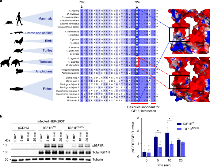
expands lifespan, especially in response to caloric restriction48,49. Finally, we have annotated a specific variant in IGF1R that is expected to affect the interaction between this receptor and the IGF1/2 growth factors50. Notably, a homology model of this region in IGF1R in C. abingdonii
suggests that position 724 is located at the surface of the protein,
and the presence of an aspartic acid residue changes the local
electrostatic field (Fig. 4a). The extended lifespan in different species correlates with IGF signalling decrease51,52, which suggests that this unique change in IGF1R
may provide an attractive target to study the cellular mechanisms
underlying the exceptional lifespan of these animals. To explore the
functional consequences of differential IGF1 signalling caused by the
p.N724D variant found in the IGF1 receptor (IGF1R), we infected HEK-293T
cells with pCDH, pCDH-IGF1RWT and pCDH-IGF1RN724D
plasmids.
Cells expressing the mutant receptor showed an attenuation of
IGF1 signalling, compared with those expressing the wild-type protein,
measured as a significant reduction in the phosphorylation levels of
IGF1R at 5 min (95% confidence interval of difference: 0.1119–1.5330, t = 2.454, P = 0.026) and 10 min (95% confidence interval of difference: 0.1991–1.6200, t = 2.714, P = 0.0153) after IGF1 treatment (Fig. 4b, Supplementary Section 7.6.2 and Supplementary Fig. 31). According to a two-way analysis of variance, the exogenous IGF1R form accounted for 16.07% of total variation (F1,4 = 20.91, P = 0.0102), while time accounted for 44.23% of total variation (F3,12 = 6.57, P = 0.0071). Interestingly, we also found in tortoises a short deletion in the coding region of IGF2R that results in the loss of two amino acids. The fact that IGF2R variants have been associated with human longevity53
opens the possibility that the variant found in tortoises could also
contribute to increasing the lifespan of these long-lived animals.
Fig. 4: Functional relevance of IGF1RN724D in the IGF1 signalling pathway.
a, Alignment of IGF1R around residue p.N724 in C. abingdonii, A. gigantea and other representative species. The predicted electrostatic surfaces of human (top right) and modelled C. abingdonii
(bottom right) IGF1R around the same residue are shown for comparison.
Negatively charged areas are depicted in red, while positively charged
areas are depicted in blue. b, Western blot analysis and
densitometry quantification of the phospho-IGF1R (pIGF1R)/total IGF1R
ratio at 5, 10 and 20 min intervals after IGF1 addition in HEK-293T
cells infected with pCDH, pCDH-IGF1RWT and pCDH-IGF1RN724D plasmids. Bars indicate means ± s.e.m. *P < 0.05, Fisher’s least significant difference test (n = 3 independent experiments).
In
summary, in this work, we report the preliminary characterization of
giant tortoise genomes. We complemented the automatic annotation of
genomes from two giant tortoise species with a hypothesis-driven
strategy using manually supervised annotation of a large set of genes.
The analysis of the resulting sequences offers candidate genes and
pathways that may underlie the extraordinary characteristics of these
iconic species, including their development, gigantism and longevity. A
better understanding of the processes that we have studied may help to
further elucidate the biology of these species and therefore aid the
ongoing efforts to conserve these dwindling lineages. Lonesome
George—the last representative of C. abingdonii, and a renowned
emblem of the plight of endangered species—left a legacy including a
story written in his genome whose unveiling has just started.
Methods
Genome sequencing and assembly
We obtained DNA from a blood sample from Lonesome George—the last member of C. abingdonii.
This DNA was sequenced, using the Illumina HiSeq 2000 platform, from a
180-base pair-insert paired-end library, a 5-kilobase (kb)-insert
mate-pair library and a 20-kb-insert mate-pair library. These libraries
were assembled with the AllPaths algorithm54
for a draft genome containing 64,657 contigs with an N50 of 74 kb.
Then, we scaffolded the contigs with SSPACE version 3.0 (ref. 55) using the long-insert mate-pair libraries. Finally, we filled the gaps with PBJelly version 15.8.24 (ref. 56)
using the reads obtained from 18 BioPac cells. This step yielded 10,623
scaffolds with an N50 of 1.27 megabases, for a final assembly
2.3 gigabases long. Then, we soft-masked repeated regions using
RepeatMasker (http://www.repeatmasker.org)
with a database containing chordate repeated elements (included in the
software) as a reference.
Additionally, we assessed the completeness of
assembly by their estimated gene content, using Benchmarking Universal
Single-Copy Orthologs (BUSCO version 3.0.0)57, which tested the status of a set of 2,586 vertebrata genes from the comprehensive catalogue of orthologues58. We also performed RNA-Seq from C. abingdonii blood and A. gigantea granuloma, and aligned the resulting reads to the assembled genome using TopHat59 (version 2.0.14). Finally, we obtained whole-genome data from A. gigantea with one Illumina lane of a 180-base pair paired-end library. The resulting reads were aligned to the C. abingdonii genome with BWA60 (version 0.7.5a). Raw reads from C. abingdonii
were also aligned to the genome for manual curation of the results. All
work on field samples was conducted at Yale University under
Institutional Animal Care and Use Committee permit number 2016-10825,
Galapagos Park Permit PC-75-16 and Convention on International Trade in
Endangered Species number 15US209142/9.
Genome annotation
Using the genome assembly of C. abingdonii and the RNA-Seq reads from C. abingdonii and A. gigantea, we performed de novo annotation with MAKER2. The algorithm was also fed both human and P. sinensis reference sequences, and performed two runs in a Microsoft Azure virtual machine (Supplementary Table 16).
In parallel, we used selected genes from the human protein database in
Ensembl as a reference to manually predict the corresponding homologues
in the genome of C. abingdonii using the BATI algorithm (Blast, Annotate, Tune, Iterate)61.
Briefly, this algorithm allows a user to annotate the position and
intron/exon boundaries of genes in novel genomes from tblastn results.
In addition, tblastn results are integrated to search for novel
homologues in the explored genome. Sequencing data have been deposited
at the Sequence Read Archive (https://www.ncbi.nlm.nih.gov/sra), with comments showing which regions were filled with the BioPac reads and therefore may contain frequent errors.
Effective population size changes and diversity
We reconstructed changes in the effective population over time using the PSMC model5
in the following way: the reads of both individuals were aligned to the
reference assembly using bwa mem (version 0.7.15-r1140). We then
constructed pseudodiploid sequences using variant calls generated with
SAMtools and BCFtools62,
requiring minimal base and mapping qualities of 30. We additionally
masked out any region with coverage below 36 or above 216 for the C. abingdonii sample, and below 8 or above 52 for the A. gigantea
sample, as a function of their respective genome-wide average coverage.
The resulting sequences were used to run 100 PSMC bootstrap replicates
per individual, using the following parameters: -N25 -t15 -r5 -p
‘4 + 25*2 + 4 + 6’. The result was averaged and scaled to real time
assuming a mutation rate (μ) of 2.5 × 10⁻⁸ and a generation time (g) of 25 years.
Expansion of gene families
To
detect expansion of gene families, we aligned pairwise all the
predicted proteins from the automatic annotation to the UniProt63 database of human proteins and the UniProt database of P. sinensis proteins using BLAST64
(version 2.6.011). Then, we used in-house Perl scripts to group these
proteins in one-to-one, one-to-many and many-to-many orthologous
relationships. Only alignments spanning at least 80% of the longer
protein, and with more than 60% identities, were considered. Finally, we
interrogated the resulting database to find families with C. abingdonii-specific
expansions and curated the results manually. This way, we constructed
extended orthology sets that may contain more than one sequence per
species. These sets recapitulate most of the known families, although
some of these families appear split according to sequence similarity.
Phylogenetic, evolutionary and structural analyses
Next,
we assessed evidence for signatures of positive selection affecting the
predicted set of genes. For this purpose, we used databases from the
human (Homo sapiens), mouse (Mus musculus), dog (Canis lupus familiaris), gecko (Gekko japonicus), green anole lizard (A. carolinensis), python snake (Python bivittatus), common garter snake (Thamnophis sirtalis), Habu viper (Trimeresurus mucrosquamatus), budgerigar (Melopsittacus undulatus), zebra finch (Taeniopygia guttata), flycatcher (Ficedula albicollis), duck (Anas platyrhynchos), turkey (Meleagris gallopavo), chicken (Gallus gallus), Chinese soft-shell turtle (P. sinensis), green sea turtle (Chelonia mydas) and painted turtle (C. picta bellii)
to generate pairwise alignments of all available genes one by one. To
this end, we used BLAST and simple in-house Perl scripts (https://github.com/vqf/LG),
which allowed us to group the genes by identity (focusing only on those
presenting one-to-one orthology). We then discarded those groups in
which there were more than three species missing (always excluding those
in which C. abingdonii was missing).
This way, we obtained 1,592
groups of sequences (similar to other studies). We then aligned them
with PRANK version 150803 using the codon model and analysed the
alignments with codeml from the PAML package65. To search for genes with signatures of positive selection affecting genes specific to C. abingdonii, we executed two different branch models—M0, with a single ω0 value (where ω represents the ratio of non-synonymous to synonymous substitutions) for all the branches (nested), and M2a, with a foreground ω2 value exclusive for C. abingdonii and a background ω1 value for all the other branches.
As a control, the second model was repeated using P. sinensis as the foreground branch. Genes with a high ω2 value (>1) and a low ω1 value (ω1 < 0.2 and ω1 ~ ω0) in C. abingdonii, but not in P. sinensis (Supplementary Section 1.2 and Supplementary Tables 5 and 17),
were then considered to be under positive selection. After this, we
used the M8 model to assess the individual importance of every site in
these positively selected genes, obtaining a list of sites of special
interest in this evolutionary effect. These results were compared with
those of the Aldabra tortoise through alignments, to evaluate which of
these important residues were altered (Supplementary Table 18). Homology models were performed with SWISS-MODEL66
from the closest template available. The results were inspected and
rendered with DeepView version 4.0.1. Electric potentials were
calculated with DeepView using the Poisson–Boltzmann computation method.
Figures were generated with PovRay (http://povray.org).
Functional analyses
HEK-293T
cells were infected with pCDH, pCDH-NEIL1, pCDH-RMI2 or
pCDH-NEIL1 + pCDH-RMI2 in the case of repair studies, and pCDH,
pCDH-IGF1RWT or pCDH-IGF1RN724D in the case of
IGF1R analyses. For the repair studies, we isolated clones of infected
HEK-293T cells with proper expression levels of NEIL1 and RMI2. Cells were exposed to ultraviolet light (20 J m−2) or H2O2
(500 μM) 24 and 48 h before being lysed in NP-40 lysis buffer
containing 50 mM Tris-HCl pH 7.4, 150 mM NaCl, 10 mM EDTA pH 8 and 1%
NP-40, and supplemented with protease inhibitor cocktail (cOmplete,
EDTA-free; Roche), as well as phosphatase inhibitors (PhosSTOP;
Roche/NaF; Merck). For the IGF1R variant analyses, cells were
serum starved for 14 h, then treated with 100 nM IGF1 for 5, 10 and
20 min before lysis in the same buffer. Equal amounts of protein were
resolved by 8 to 13% sodium dodecyl sulfate polyacrylamide gel
electrophoresis and transferred to PVDF membranes (GE Healthcare Life
Sciences). Membranes were blocked for 1 h at room temperature with TBS-T
(0.1% Tween 20) containing 5% bovine serum albumin. Immunoblotting was
performed with primary antibodies diluted 1:500 to 1:1000 in TBS-T and
1% bovine serum albumin and incubated overnight at 4 °C. The primary
antibodies used were: anti-phospho-Histone H2AX (Ser139) (EMD Millipore;
05-636, clone JBW301, lot 2854120), anti-PARP (Cell Signaling
Technology; 9542S, rabbit polyclonal, lot 15), anti-FLAG (Cell Signaling
Technology; 2368S, rabbit polyclonal, lot 12), anti-IGF1R (Abcam;
ab182408, clone EPR19322, lot GR312678-8), anti-IGF1R (p Tyr1161) (Novus
Biologicals; NB100-92555, rabbit polyclonal, lot CJ36131), anti-β-actin
(Sigma–Aldrich, A5441, clone AC-15, lot 014M4759) and anti-α-tubulin
(Sigma–Aldrich, T6074, clone B-5-1-2, lot 075M4823V). After washing with
TBS-T, membranes were incubated with secondary antibodies conjugated
with IRDye 680RD (LI-COR Biosciences; 926-68071, polyclonal
goat-anti-rabbit, lot C41217-03; and 926-32220, polyclonal
goat-anti-mouse, lot C00727-03) or IRDye 800CW (LI-COR Biosciences;
926-32211, polyclonal goat-anti-rabbit, lot C60113-05; and 926-32210,
polyclonal goat-anti-mouse, lot C50316-03) for 1 h at room temperature.
Protein bands were scanned on an Odyssey infrared scanner (LI-COR
Biosciences). Band intensities were quantified by ImageJ and used to
calculate the phospho-IGF1R/IGF1R ratio in the case of the IGF1R assay.
In each replicate, cells were infected independently. For the samples
from ultraviolet treatment, Flag (RMI2) was detected on the same samples
used for the remaining western blots shown in this panel, run in
parallel on an identical blot. Similarly, for the samples from H2O2
treatment, the western blots shown were carried out with the same
samples run in parallel in three identical blots (one for PARP and
actin, a second for Flag (NEIL1 and RMI2) and a third for pH2AX). Each
sample contained one replicate. Statistical comparisons consisted of
two-way analysis of variance performed using GraphPad Prism 7.0
software. Differences were considered statistically significant when P < 0.05. Effect sizes are expressed as group sum-of-squares divided by the total sum-of-squares (R2). At each time point, both groups were also compared with Fisher’s least significant difference test (uncorrected; α = 0.05).
Kehlmaier, C. et al. Tropical ancient DNA reveals relationships of the extinct Bahamian giant tortoise Chelonoidis alburyorum. Proc. R. Soc. B284, 20162235 (2017).
Campbell, M. S., Holt, C., Moore, B. & Yandell, M. Genome annotation and curation using MAKER and MAKER‐P. Curr. Protoc. Bioinformatics48, 1–39 (2014).
Wang,
Z. et al. The draft genomes of soft-shell turtle and green sea turtle
yield insights into the development and evolution of the turtle-specific
body plan. Nat. Genet.45, 701–706 (2013).
Sanchis-Gomar,
F. et al. A preliminary candidate approach identifies the combination
of chemerin, fetuin-A, and fibroblast growth factors 19 and 21 as a
potential biomarker panel of successful aging. Age37, 9776 (2015).
Van
der Goot, A. T. et al. Delaying aging and the aging-associated decline
in protein homeostasis by inhibition of tryptophan degradation. Proc. Natl Acad. Sci. USA109, 14912–14917 (2012).
Chiari,
Y., Cahais, V., Galtier, N. & Delsuc, F. Phylogenomic analyses
support the position of turtles as the sister group of birds and
crocodiles (Archosauria). BMC Biol.10, 65 (2012).
Li,
Y. I., Kong, L., Ponting, C. P. & Haerty, W. Rapid evolution of
beta-keratin genes contribute to phenotypic differences that distinguish
turtles and birds from other reptiles. Genome Biol. Evol.5, 923–933 (2013).
Barreiro,
L. B. & Quintana-Murci, L. From evolutionary genetics to human
immunology: how selection shapes host defence genes. Nat. Rev. Genet.11, 17–30 (2010).
Zimmerman,
L. M., Vogel, L. A. & Bowden, R. M. Understanding the vertebrate
immune system: insights from the reptilian perspective. J. Exp. Biol.213, 661–671 (2010).
Voskoboinik, I., Whisstock, J. C. & Trapani, J. A. Perforin and granzymes: function, dysfunction and human pathology. Nat. Rev. Immunol.15, 388–400 (2015).
Jaffe,
A. L., Slater, G. J. & Alfaro, M. E. The evolution of island
gigantism and body size variation in tortoises and turtles. Biol. Lett.7, 558–561 (2011).
Chuang,
D. M., Hough, C. & Senatorov, V. V. Glyceraldehyde-3-phosphate
dehydrogenase, apoptosis, and neurodegenerative diseases. Annu. Rev. Pharmacol. Toxicol.45, 269–290 (2005).
Corti, P. et al. Globin X is a six-coordinate globin that reduces nitrite to nitric oxide in fish red blood cells. Proc. Natl Acad. Sci. USA113, 8538–8543 (2016).
Schwarze,
K., Singh, A. & Burmester, T. The full globin repertoire of turtles
provides insights into vertebrate globin evolution and functions. Genome Biol. Evol.7, 1896–1913 (2015).
Zhao, Y. et al. Codon 104 variation of p53 gene provides adaptive apoptotic responses to extreme environments in mammals of the Tibet plateau. Proc. Natl Acad. Sci. USA110, 20639–20644 (2013).
Chiari,
Y., Glaberman, S. & Lynch, V. J. Insights on cancer resistance in
vertebrates: reptiles as a parallel system to mammals. Nat. Rev. Cancer18, 525 (2018).
Garner,
M. M., Hernandez-Divers, S. M. & Raymond, J. T. Reptile neoplasia: a
retrospective study of case submissions to a specialty diagnostic
service. Vet. Clin. North Am. Exot. Anim. Pract.7, 653–671 (2004).
Martinvalet,
D., Zhu, P. & Lieberman, J. Granzyme A induces caspase-independent
mitochondrial damage, a required first step for apoptosis. Immunity22, 355–370 (2005).
Gorbunova,
V., Seluanov, A., Zhang, Z., Gladyshev, V. N. & Vijg, J.
Comparative genetics of longevity and cancer: insights from long-lived
rodents. Nat. Rev. Genet.15, 531–540 (2014).
Daley,
J. M., Chiba, T., Xue, X., Niu, H. & Sung, P. Multifaceted role of
the Topo III⍺–RMI1-RMI2 complex and DNA2 in the BLM-dependent pathway of
DNA break end resection. Nucleic Acids Res.42, 11083–11091 (2014).
Ivashkevich,
A., Redon, C. E., Nakamura, A. J., Martin, R. F. & Martin, O. A.
Use of the gamma-H2AX assay to monitor DNA damage and repair in
translational cancer research. Cancer Lett.327, 123–133 (2012).
Cremona,
C. A. et al. Extensive DNA damage-induced sumoylation contributes to
replication and repair and acts in addition to the mec1 checkpoint. Mol. Cell45, 422–432 (2012).
Wang, Y., Ghosh, G. & Hendrickson, E. A. Ku86 represses lethal telomere deletion events in human somatic cells. Proc. Natl Acad. Sci. USA106, 12430–12435 (2009).
Ribes-Zamora,
A., Indiviglio, S. M., Mihalek, I., Williams, C. L. & Bertuch, A.
A. TRF2 interaction with Ku heterotetramerization interface gives
insight into c-NHEJ prevention at human telomeres. Cell Rep.5, 194–206 (2013).
Shikama, N., Ackermann, R. & Brack, C. Protein synthesis elongation factor EF-1 alpha expression and longevity in Drosophila melanogaster. Proc. Natl Acad. Sci. USA91, 4199–4203 (1994).
Ohta, S., Ohsawa, I., Kamino, K., Ando, F. & Shimokata, H. Mitochondrial ALDH2 deficiency as an oxidative stress. Ann. NY Acad. Sci.1011, 36–44 (2004).
Serizawa,
A., Dando, P. M. & Barrett, A. J. Characterization of a
mitochondrial metallopeptidase reveals neurolysin as a homologue of
thimet oligopeptidase. J. Biol. Chem.270, 2092–2098 (1995).
Tristan,
C., Shahani, N., Sedlak, T. W. & Sawa, A. The diverse functions of
GAPDH: views from different subcellular compartments. Cell. Signal.23, 317–323 (2011).
Fan,
C. et al. MIF intersubunit disulfide mutant antagonist supports
activation of CD74 by endogenous MIF trimer at physiologic
concentrations. Proc. Natl Acad. Sci. USA110, 10994–10999 (2013).
Verschuren,
L. et al. MIF deficiency reduces chronic inflammation in white adipose
tissue and impairs the development of insulin resistance, glucose
intolerance, and associated atherosclerotic disease. Circ. Res.105, 99–107 (2009).
Harper,
J. M., Wilkinson, J. E. & Miller, R. A. Macrophage migration
inhibitory factor-knockout mice are long lived and respond to caloric
restriction. FASEB J.24, 2436–2442 (2010).
Whittaker, J. et al. Alanine scanning mutagenesis of a type 1 insulin-like growth factor receptor ligand binding site. J. Biol. Chem.276, 43980–43986 (2001).
Brohus,
M., Gorbunova, V., Faulkes, C. G., Overgaard, M. T. & Conover, C.
A. The insulin-like growth factor system in the long-lived naked
mole-rat. PLoS ONE10, e0145587 (2015).
Soerensen,
M. et al. Human longevity and variation in GH/IGF-1/insulin signaling,
DNA damage signaling and repair and pro/antioxidant pathway genes: cross
sectional and longitudinal studies. Exp. Gerontol.47, 379–387 (2012).
Gnerre, S. et al. High-quality draft assemblies of mammalian genomes from massively parallel sequence data. Proc. Natl Acad. Sci. USA108, 1513–1518 (2011).
Simao,
F. A., Waterhouse, R. M., Ioannidis, P., Kriventseva, E. V. &
Zdobnov, E. M. BUSCO: assessing genome assembly and annotation
completeness with single-copy orthologs. Bioinformatics31, 3210–3212 (2015).
Zdobnov,
E. M. et al. OrthoDBv9.1: cataloging evolutionary and functional
annotations for animal, fungal, plant, archaeal, bacterial and viral
orthologs. Nucleic Acids Res.45, D744–D749 (2017).
Quesada,
V., Velasco, G., Puente, X. S., Warren, W. C. & López-Otín, C.
Comparative genomic analysis of the zebra finch degradome provides new
insights into evolution of proteases in birds and mammals. BMC Genomics11, 220 (2010).
Biasini, M. et al. SWISS-MODEL: modelling protein tertiary and quaternary structure using evolutionary information. Nucleic Acids Res.42, W252–W258 (2014).
We
thank J. R. Obeso for support, J. M. Freije, X. S. Puente, R.
Valdés-Mas, F. G. Osorio, D. López-Velasco, A. Corrales, P. Salinas, D.
Rodríguez, A. López-Soto, A. R. Folgueras and M. Mittelbrunn for helpful
comments and advice, M. Garaña, O. Sanz, J. Isla and A. Marcos
(Microsoft) for computing facilities, and F. Rodríguez, D. A. Puente and
S. A. Miranda for excellent technical assistance. We also acknowledge
generous support from J. I. Cabrera. We thank Banco Santander for
funding a short stay of S.F.-R. and D.C.-I. at Yale University. V.Q. is
supported by grants from the Principado de Asturias and Ministerio de
Economía y Competitividad, including FEDER funding. L.F.K.K. is
supported by an FPI fellowship associated with BFU2014-55090-P (FEDER).
T.M.-B. is supported by MINECO BFU2017-86471-P (MINECO/FEDER, UE), an
NIH U01 MH106874 grant, the Howard Hughes International Early Career
programme, Obra Social ‘La Caixa’ and Secretaria d’Universitats i
Recerca, and CERCA Programme del Departament d’Economia i Coneixement de
la Generalitat de Catalunya. C.L.-O. is supported by grants from the
European Research Council (DeAge; ERC Advanced Grant), Ministerio de
Economía y Competitividad, Instituto de Salud Carlos III (RTICC) and
Progeria Research Foundation. The Instituto Universitario de Oncología
is supported by Fundación Bancaria Caja de Ahorros de Asturias. We also
thank staff at the Galapagos National Park and Galapagos Conservancy for
logistic and financial support.
Author information
Author notes
These authors contributed equally: Víctor Quesada, Sandra Freitas-Rodríguez, Joshua Miller, José G. Pérez-Silva.
Affiliations
Departamento
de Bioquímica y Biología Molecular, Instituto Universitario de
Oncología del Principado de Asturias, CIBERONC, Universidad de Oviedo,
Oviedo, Spain
Víctor Quesada
, Sandra Freitas-Rodríguez
, José G. Pérez-Silva
, Olaya Santiago-Fernández
, Diana Campos-Iglesias
, Miguel G. Álvarez
, Dido Carrero
, Miguel Araujo-Voces
, Pablo Mayoral
, Javier R. Arango
, Isaac Tamargo-Gómez
, David Roiz-Valle
, María Pascual-Torner
, Gabriel Bretones
& Carlos López-Otín
Department of Ecology and Evolutionary Biology, Yale University, New Haven, CT, USA
Joshua Miller
, Maud Quinzin
, Benjamin R. Evans
, Stephen J. Gaughran
& Adalgisa Caccone
Institute for Genomics and Systems Biology, The University of Chicago, Chicago, IL, USA
Zi-Feng Jiang
& Kevin P. White
Galapagos National Park Directorate, Galapagos Islands, Ecuador
Washington Tapia
& Danny O. Rueda
Galapagos Conservancy, Fairfax, VA, USA
Washington Tapia
Institute of Evolutionary Biology (UPF-CSIC), Barcelona, Spain
Lukas F. K. Kuderna
& Tomàs Marquès-Bonet
CNAG-CRG, Centre for Genomic Regulation, Barcelona Institute of Science and Technology , Barcelona, Spain
Lukas F. K. Kuderna
& Tomàs Marquès-Bonet
College of Science and Engineering, Flinders University, Adelaide, South Australia, Australia
Luciano B. Beheregaray
College of Environmental Science and Forestry, State University of New York, Syracuse, NY, USA
James P. Gibbs
Department of Biology, University of South Alabama, Mobile, AL, USA
Ylenia Chiari
& Scott Glaberman
Department of Biology, University of Florence, Florence, Italy
Claudio Ciofi
School of Natural Sciences, University of California, Merced, CA, USA
Danielle L. Edwards
Department of Biology, University of Mississippi, Oxford, MS, USA
Ryan C. Garrick
Department of Biology, The University of British Columbia, Kelowna, British Columbia, Canada
Michael A. Russello
Department of Biology, School of Sciences and Engineering, University of Crete, Heraklion, Greece
Nikos Poulakakis
Natural History Museum of Crete, Heraklion, Greece
Nikos Poulakakis
Catalan Institution of Research and Advanced Studies, Barcelona, Spain
Tomàs Marquès-Bonet
Institut Català de Paleontologia Miquel Crusafont, Universitat Autònoma de Barcelona, Barcelona, Spain
Tomàs Marquès-Bonet
Contributions
V.Q.
and J.G.P.-S. performed the automatic analysis of genomes. S.F.-R.
coordinated the manual genomic annotation, which was performed by
J.G.P.-S., O.S.-F., D.C.-I., M.G.A., M.A.-V., D.C., P.M., J.R.A.,
I.T.-G., D.R.-V. and M.P.-T. S.F.-R. and D.C.-I. performed the
validation of the identified genomic variants. G.B. coordinated the
functional analyses of the identified genomic variants, which were
carried out by O.S.-F., D.C.-I., M.G.A., M.A.-V., D.C., P.M., J.R.A. and
I.T.-G. J.M. helped to screen the wild samples for SNP validation, and
contributed to results interpretation. M.Q., L.B.B., J.P.G., Y.C., S.G.,
C.C., B.R.E., S.J.G., D.L.E., R.C.G., M.A.R. and N.P. contributed to
early data collection and analyses. W.T., D.O.R. and J.P.G. helped to
obtain material-securing permits and biological samples. K.P.W. partly
supported data collection and supervised the initial analysis. Z.-F.J.
prepared DNA and RNA samples for genomic analyses and conducted raw data
quality checks. L.F.K.K. and T.M.-B. performed population history and
diversity studies. V.Q., A.C. and C.L.-O. directed the research,
analysed the data and wrote the manuscript.
Table S16. Summary of average characteristics of the automatic annotation set of C. abingdonii compared to sets of other species. Table S17. Genes with signatures of positive selection in C. abingdonii. Table S18. Positions of interest within the genes with signatures of positive selection. Table S19. microRNAs identified in C. abingdonii's genome. Table S20. Primers used for validation of gene alterations identified in C. abingdonii's
genome. Table S21. Expansion events with coverage and identity for
nucleotide and amino acid sequences compared to one of the paralogues
Nenhum comentário:
Postar um comentário
Observação: somente um membro deste blog pode postar um comentário.
Nenhum comentário:
Postar um comentário
Observação: somente um membro deste blog pode postar um comentário.