Synchrotron X-ray sheds light on some of the world's oldest dinosaur eggs
Raio-X síncrotron lança luz sobre alguns dos ovos de dinossauro mais antigos do mundo
An international team of scientists led by the University of
the Witwatersrand in South Africa has produced highly detailed
reproductions of the skulls of some of the oldest known dinosaur embryos
in 3-D, using powerful and non-destructive synchrotron techniques at
the ESRF, the European Synchrotron in France. They found that the skulls
develop in the same order as those of today's crocodiles, chickens,
turtles and lizards. The findings are published today in Scientific Reports.
The scientists have published the 3-D reconstructions of the ~2-cm-long embryonic skulls in Scientific Reports.
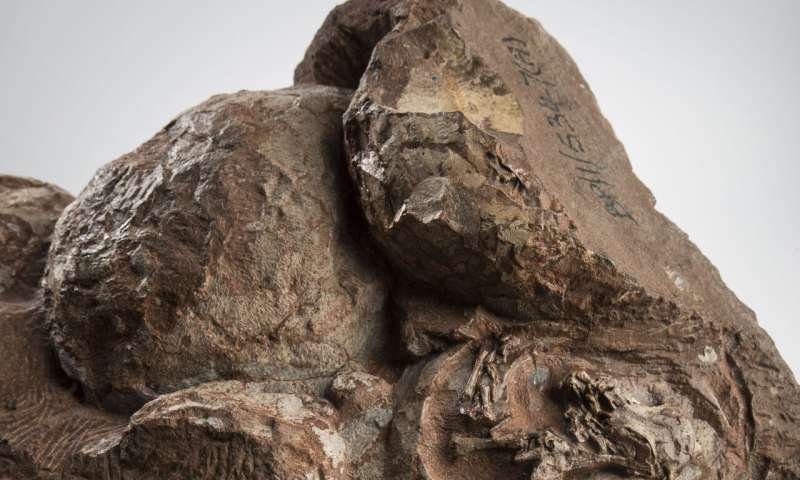
The embryos, found in 1976 in Golden Gate Highlands National Park (Free
State Province, South Africa) belong to South Africa's iconic dinosaur
Massospondylus carinatus, a 5-meter-long herbivore that nested in the
Free State region 200 million years ago.
The scientific usefulness of the embryos was previously limited by
their extremely fragile nature and tiny size. In 2015, scientists Kimi
Chapelle and Jonah Choiniere from the University of Witwatersrand
brought them to the European Synchrotron (ESRF) in Grenoble, France for
scanning. At the ESRF, an 844-metre ring of electrons traveling at the
speed of light emits high-powered X-ray beams that can be used to scan
matter non-destructively, including fossils. The embryos were scanned at
an unprecedented level of detail, at the resolution of an individual
bone cell.
With these data in hand, and after nearly three years of data
processing at the Wits laboratory, the team was able to reconstruct a
3-D model of the baby dinosaur skull. "No lab CT scanner in the world
can generate these kinds of data," said Vincent Fernandez, one of the
co-authors and scientist at the Natural History Museum in London (U.K.).
"Only with a huge facility like the ESRF can we unlock the hidden
potential of our most exciting fossils. This research is a great example
of a global collaboration between Europe and the South African National
Research Foundation," he adds.
------------------------------------------------------------------------------
Uma equipe internacional de cientistas liderada pela Universidade de Witwatersrand, na África do Sul, produziu reproduções altamente detalhadas dos crânios de alguns dos mais antigos embriões de dinossauros conhecidos em 3D, usando técnicas síncrotron poderosas e não destrutivas no ESRF, o European Síncrotron na França. Eles descobriram que os crânios se desenvolvem na mesma ordem que os crocodilos, galinhas, tartarugas e lagartos de hoje. Os resultados são publicados hoje no Scientific Reports.
Os cientistas publicaram as reconstruções em 3D dos crânios embrionários de 2 cm de comprimento no Scientific Reports. Os embriões, encontrados em 1976 no Parque Nacional Golden Gate Highlands (Província de Estado Livre, África do Sul) pertencem ao icônico dinossauro da África do Sul Massospondylus carinatus, um herbívoro de 5 metros de comprimento e aninhado na região de Estado Livre há 200 milhões de anos.
A utilidade científica dos embriões era anteriormente limitada por sua natureza extremamente frágil e tamanho pequeno. Em 2015, os cientistas Kimi Chapelle e Jonah Choiniere, da Universidade de Witwatersrand, os trouxeram ao Síncrotron Europeu (ESRF) em Grenoble, na França, para digitalização. No ESRF, um anel de elétrons de 844 metros que viaja à velocidade da luz emite feixes de raios X de alta potência que podem ser usados para varrer a matéria de maneira não destrutiva, incluindo fósseis. Os embriões foram escaneados com um nível de detalhe sem precedentes, na resolução de uma célula óssea individual.
Com esses dados em mãos, e após quase três anos de processamento de dados no laboratório Wits, a equipe conseguiu reconstruir um modelo 3D do crânio do bebê dinossauro. "Nenhum scanner de laboratório no mundo pode gerar esses tipos de dados", disse Vincent Fernandez, um dos co-autores e cientista do Museu de História Natural de Londres (Reino Unido). "Somente com uma enorme instalação como a ESRF podemos desbloquear o potencial oculto de nossos fósseis mais emocionantes. Esta pesquisa é um ótimo exemplo de uma colaboração global entre a Europa e a Fundação Nacional de Pesquisa da África do Sul", acrescenta ele.
-------------------------------------------------------
Up until now, it was believed that the embryos in those eggs had died
just before hatching. However, during the study, lead author Chapelle
noticed similarities with the developing embryos of living dinosaur
relatives (crocodiles, chickens, turtles, and lizards). By comparing
which bones of the skull were present at different stages of their
embryonic development, Chapelle and co-authors demonstrated that the
Massospondylus embryos were actually much younger than previously
thought and were only at 60% through their incubation period.
The team also found that each embryo had two types of teeth preserved
in its developing jaws. One set was made up of very simple triangular
teeth that would have been resorbed or shed before hatching, just like
geckos and crocodiles today. The second set was very similar to those of
adults, and would be the ones that the embryos hatched with. "I was
really surprised to find that these embryos not only had teeth, but had
two types of teeth. The teeth are so tiny; they range from 0.4 to 0.7 mm
wide. That's smaller than the tip of a toothpick," says Chapelle.
The researchers concluded that the dinosaurs
developed in the egg just like their reptilian relatives, whose
embryonic developmental pattern hasn't changed in 200 million years.
"It's incredible that in more than 250 million years of reptile
evolution, the way the skull develops in the egg remains more or less
the same. Goes to show—you don't mess with a good thing," says Jonah
Choiniere, professor at the University of Witwatersrand and also
co-author of the study.
The team hopes to apply their method to other dinosaur embryos to
estimate their level of development. They will be looking at the rest of
the skeleton of the Massospondylus embryos to see if it also shares
similarities in development with today's dinosaur relatives. The arms
and legs of the Massospondylus embryos have already been used to show that hatchlings likely walked on two legs.
More information:
Conserved in-ovo cranial ossification sequences of extant
saurians allow estimation of embryonic dinosaur developmental stages, Scientific Reports (2020).
Kimberley E. J. Chapelle et al. A quantitative method for inferring
locomotory shifts in amniotes during ontogeny, its application to
dinosaurs and its bearing on the evolution of posture, Palaeontology (2019). DOI: 10.1111/pala.12451
Nenhum comentário:
Postar um comentário
Observação: somente um membro deste blog pode postar um comentário.