quinta-feira, 30 de setembro de 2010

Água doce e ameaçada

30/9/2010

Agência FAPESP – Uma análise em escala global, que poderá auxiliar a identificar as áreas em que a disponibilidade de água para consumo estão mais ameaçadas no mundo, é o destaque na edição desta quinta-feira (30/9) da revista Nature.

A pesquisa, feita por cientistas da Austrália, Estados Unidos, Alemanha e China, destaca as maiores ameaças tanto à segurança da água como à biodiversidade nos rios do planeta.
Apesar de a água ser o mais essencial dos recursos naturais, os sistemas de água doce que podem ser usados para consumo humano estão fortemente ameaçados justamente pelo homem, principalmente pela poluição e também como resultado de processos como a urbanização, industrialização, irrigação e a construção de reservatórios.
Soma-se a isso o aquecimento global e o resultado está longe de ser animador. Segundo os autores do estudo, desenvolver alternativas que possam reverter essa tendência exige primeiramente que se tenha um diagnóstico das ameaças à segurança da água tanto no nível local como no global.

Charles Vorosmarty, da Universidade da Cidade de Nova York, e colegas descrevem no artigo um modelo espacial que fornece uma análise global das ameaças à água doce. É o primeiro modelo a considerar a segurança da água usada para consumo humano em conjunto com a biodiversidade aquática.
Além de listar quais são as principais ameaças, o estudo identifica as áreas de maior risco. Segundo o trabalho, grandes investimentos no uso e no tratamento de água nos países mais ricos têm beneficiado seus habitantes, diminuindo em até 95% a ameaça à segurança da água nesses locais.
Em compensação, nos países mais pobres os investimentos reduzidos no setor implicam que a vulnerabilidade ao problema nessas áreas é muito elevada.
Mas, mesmo nas nações mais ricas, de acordo com a pesquisa, os investimentos em massa em tecnologia de uso da água têm permitido “ofuscar os níveis de estresse”, mas sem remediar as causas por trás do problema.
O estudo concluiu que 80% da população mundial está exposta a níveis elevados de ameaças à segurança da água. A falta de investimento no cuidado dos rios e corpos de água doce também coloca em risco a biodiversidade, indicam os autores.
“A proliferação de áreas densamente povoadas em zonas costeiras, incluindo megacidades, implica que seus rios apresentem alto nível de ameaças à segurança da água por virtualmente toda a sua extensão, como, por exemplo, o Paraíba do Sul, no Estado de São Paulo, o Pasig, em Manila, nas Filipinas, e o Ogun, em Lagos, na Nigéria”, disseram.
Segundo eles, é urgente a necessidade de mobilizar recursos para apoiar abordagens que possam enfrentar o problema. “Sem grandes compromissos políticos e financeiros, contrastes marcantes na segurança da água usada por humanos continuarão a separar os ricos dos pobres”, afirmam.
O artigo Global threats to human water security and river biodiversity (doi:10.1038/nature09440), de Charles Vorosmarty e outros, pode ser lido por assinantes da Nature em www.nature.com.

Fonte: Fapesp

Contaminantes emergentes na água

30/9/2010

Por Fabio Reynol
 
Agência FAPESP – Durante a década de 1990, houve uma redução na população de jacarés que habitava os pântanos da Flórida, nos Estados Unidos. Ao investigar o problema, cientistas perceberam que os machos da espécie tinham pênis menores do que o normal, além de apresentar baixos índices do hormônio masculino testosterona.
Os estudos verificaram que as mudanças hormonais que estavam alterando o fenótipo dos animais e prejudicando sua reprodução foram desencadeadas por pesticidas clorados empregados em plantações naquela região.
Esses produtos químicos eram aplicados de acordo com a legislação norte-americana, a qual estabelecia limites máximos baseados em sua toxicidade, mas não considerava a alteração hormonal que eles provocavam, simplesmente porque os efeitos não eram conhecidos.
Assim como os pântanos da Flórida, corpos d’água de vários pontos do planeta estão sendo contaminados com diferentes coquetéis que podem conter princípios ativos de medicamentos, componentes de plásticos, hormônios naturais e artificiais, antibióticos, defensivos agrícolas e muitos outros em quantidades e proporções diversas e com efeitos desconhecidos para os animais aquáticos e também para pessoas que consomem essas águas.
“Em algumas dessas áreas, meninas estão menstruando cada vez mais cedo e, nos homens, o número de espermatozoides despencou nos últimos 50 anos. Esses são alguns problemas cujos motivos ninguém conseguiu explicar até agora e que podem estar relacionados a produtos presentes na água que bagunçam o ciclo hormonal”, disse Wilson Jardim, professor titular do Instituto de Química da Universidade Estadual de Campinas (Unicamp), à Agência FAPESP.
O pesquisador conta que esses contaminantes, chamados emergentes, podem estar por trás de vários outros efeitos relacionados tanto à saúde humana como aos ecossistemas aquáticos.
“Como não são aplicados métodos de tratamento que retirem esses contaminantes, as cidades que ficam à jusante de um rio bebem o esgoto das que ficam à montante”, alertou o pesquisador que coordena o Projeto Temático “Ocorrência e atividade estrogênica de interferentes endócrinos em água para consumo humano e em mananciais do Estado de São Paulo”, apoiado pela FAPESP.
O aumento no consumo de cosméticos, de artigos de limpeza e de medicamentos tem piorado a situação, de acordo com o pesquisador, cujo grupo encontrou diversos tipos de produtos em amostras de água retirada de rios no Estado de São Paulo. O antiinflamatório diclofenaco, o analgésico ácido acetilsalicílico e o bactericida triclosan, empregado em enxaguatórios bucais, são apenas alguns exemplos.
A esses se soma uma crescente coleção de cosméticos que engorda o lixo químico que vai parar nos cursos d’água sem receber tratamento algum. “Estima-se que uma pessoa utilize, em média, dez produtos cosméticos e de higiene todos os dias antes mesmo de sair de casa”, disse Jardim.
Sem uma legislação que faça as empresas de distribuição retirar essas substâncias tanto do esgoto a ser jogado nos rios como da água deles captada, tem sido cada vez mais comum encontrar interferentes hormonais nas torneiras das residências. Os filtros domésticos disponíveis no mercado não dão conta dessa limpeza.
“Os métodos utilizados pelas estações de tratamento de água brasileiras são em geral seculares. Eles não incorporaram novas tecnologias, como a oxidação avançada, a osmose inversa e a ultrafiltração”, disse o professor da Unicamp, afirmando acreditar que tais métodos só serão incorporados pelas empresas por meio de uma legislação específica, uma vez que eles encareceriam o tratamento.
Peixes feminilizados
Uma das primeiras cidades a enfrentar esse tipo de contaminação foi Las Vegas, nos Estados Unidos. Em meio a um deserto, o município depende de uma grande quantidade de água retirada do lago Mead, o qual também recebe o esgoto da cidade.
Apesar de contar com um bom tratamento de esgoto, a água da cidade acabou provocando alterações hormonais nas comunidades de animais aquáticos do lago, com algumas espécies de peixes tendo apresentado altos índices de feminilização. Universidades e concessionárias de água se uniram para estudar o problema e chegaram à conclusão de que o esgoto precisava de melhor tratamento.
“Foi uma abordagem madura, racional e que contou com o apoio da população, que se mostrou disposta a até pagar mais em troca de uma água limpa desses contaminantes”, contou Jardim.
Alterações como o odor na água são indicadores de contaminantes como o bisfenol A, produto que está presente em diversos tipos de plásticos e que pode afetar a fertilidade, de acordo com pesquisas feitas com ratos no Instituto de Biociências do campus de Botucatu da Universidade Estadual Paulista (Unesp).
Jardim alerta que o bisfenol A é um interferente endócrino comprovado que afeta especialmente organismos em formação, o que o torna perigoso no desenvolvimento endócrino das crianças. Além dele, a equipe da Unicamp também identificou atrazina, um pesticida utilizado na agricultura.
Não apenas produtos que alteram a produção hormonal foram detectados na pesquisa, há ainda outros que afetam o ambiente e têm efeitos desconhecidos no consumo humano. Um deles é o triclosan, bactericida empregado em enxaguatórios bucais cuja capacidade biocida aumenta sob o efeito dos raios solares.
Se o efeito individual de cada um desses produtos é perigoso, pouco se sabe sobre os resultados de misturas entre eles. A interação entre diferentes químicos em proporções e quantidades inconstantes e reunidos ao acaso produz novos compostos dos quais pouco se conhecem os efeitos.
“A realidade é que não estamos expostos a cada produto individualmente, mas a uma mistura deles. Se dois compostos são interferentes endócrinos quando separados, ao juntá-los não significará, necessariamente, que eles vão se potencializar”, disse Jardim.
Segundo ele, essas interações são muito complexas. Para complicar, todos os dados de que a ciência dispõe no momento são para compostos individuais.
Superbactérias
Outra preocupação do pesquisador é a presença de antibióticos nas águas dos rios. Por meio do projeto “Antibióticos na bacia do rio Atibaia”, apoiado pela FAPESP por meio de um Auxílio à Pesquisa – Regular, Jardim e sua equipe analisaram de 2007 a 2009 a presença de antibióticos populares na água do rio paulista.
A parte da análise ficou por conta do doutorando Marco Locatelli, que identificou concentrações de cefalexina, ciprofloxacina, amoxicilina e trimetrotrin em amostras da água do Atibaia.
A automedicação e o consumo exacerbado desse tipo de medicamento foram apontados por Jardim como as principais causas dessa contaminação que apresenta como risco maior o desenvolvimento de “superbactérias”, microrganismos muito resistentes à ação desses antibióticos.
Todas essas questões foram debatidas no fim de 2009 durante o 1º Workshop sobre Contaminantes Emergentes em Águas para Consumo Humano, na Unicamp. O evento foi coordenado por Jardim e recebeu o apoio FAPESP por meio de um Auxílio à Pesquisa – Organização de Reunião Científica e/ou Tecnológica.
O professor da Unicamp reforça a gravidade da questão da água, uma vez que pode afetar de inúmeras maneiras a saúde da população e o meio ambiente. “Isso já deve estar ocorrendo de forma silenciosa e não está recebendo a devida atenção”, alertou.

Fonte: Fapesp

segunda-feira, 27 de setembro de 2010

Posted: 26 Sep 2010 08:19 PM PDT
Reconstruções dos esqueletos: Utahceratops à esquerda
© Sampson et.al.

Duas extraordinárias novas espécies de dinossauros com chifre foram encontradas no Grand Staircase-Escalante National Monument, no Sul do estado de Utah. Os gigantes herbívoros eram habitantes do "continente perdido" chamado Laramidia, formado quando um mar raso inundou a região central da América do Norte, separando as partes oeste e leste do continente por milhões de anos durante o período Cretáceo Superior. Os recentemente descobertos dinossauros, parentes próximos do famosos Triceratops, foram anunciados no site de artigos científicos, PLoS ONE, uma biblioteca de ciências pública online, cujo acesso é gratuito. Para saber mais sobre os dois novos magníficos dinossauros, acesse o resto da postagem.


Leia o resto do texto no Blog do Ikessauro: http://ikessauro.blogspot.com/

sábado, 25 de setembro de 2010

LINK PALEONTOLÓGICO

http://www.app.pan.pl/archives.html

Bivalves from Cretaceous cold−seep deposits on Hokkaido, Japan

Steffen Kiel, Kazutaka Amano, and Robert G. Jenkins

Acta Palaeontologica Polonica 53 (3), 2008: 525-537
Cretaceous cold−seep deposits of the Yezo Group on Hokkaido, Japan, yield a rich and well−preserved mollusk fauna. The systematics of nine bivalve species previously reported from these deposits can now be reevaluated using newly collected fossils. The fossils include a Cenomanian specimen of Nucinella gigantea with a drill hole possibly made by a naticid, by far the oldest record of a drill hole from a cold seep site. In Japan, Cretaceous seep bivalve assemblages are characterized by (i) the unique occurrence of large specimens of Nucinella (Manzanellidae), (ii) the commonly present nuculid Acila (Truncacila), and (iii) a high diversity of lucinids, possibly as many as four distinct genera. Two new species described are the Albian Acharax mikasaensis (Solemyidae) and the Albian to Campanian Thyasira tanabei (Thyasiridae), of which the former had previously been misidentified as the oldest vesicomyid, the latter as the oldest Conchocele.


Key words: Solemyidae, Manzanellidae, Lucinidae, Thyasiridae, hydrocarbon seeps, chemosymbiosis, Cretaceous, Japan

Steffen Kiel [steffen.kiel@gmx.de], Institut für Geowissenschaften, Christian−Albrechts−Universität Kiel, Ludewig−Meyn−Str. 10, 24118 Kiel, Germany, and Dept. of Paleobiology, Smithsonian Natural History Museum, Box 37012, Washington DC 20013−7012, USA; Kazutaka Amano [amano@juen.ac.jp], Department of Geoscience, Joetsu University of Education, Joetsu 943−8512, Japan; Robert G. Jenkins [robertgj@ynu.ac.jp], Faculty of Education and Human Sciences, Yokohama National University, Kanagawa 240−8501, Japan.

New fossil triakid sharks from the Eocene of Prémontré, France and comments on fossil record of the family

Sylvain Adnet and Henri Cappetta
Acta Palaeontologica Polonica 53 (3), 2008: 433-448
During the last two decades, an abundant selachian assemblage has been collected from the late Ypresian (NP12)
fossiliferous sands of Prémontré (Aisne, northern France) but has received little attention. Sharks of the family Triakidae (Carcharhiniformes) are particularly well represented and all are described and figured herein. Among them, two new species of the genus Galeorhinus are described: G. duchaussoisi sp. nov. and G. louisi sp. nov.; these are compared to the common Paleogene G. ypresiensis which is refigured. Another triakid taxon, the genus Gomphogaleus gen. nov., is described. Most of the triakids have been recorded elsewhere in the North Atlantic region, suggesting a wider distribution than expected for these small sharks during the Paleogene. The present paper updates the list of selachians from Prémontré, bringing the number of taxa from 19 to 33 (including 22 sharks and 11 batoids) and improving our knowledge of the ancient North Atlantic Ypresian selachian fauna. Despite this vastly improved record, it is clear that fossil data are still very incomplete and insufficient for calibrating phylogenetic hypotheses of living forms. Review of the Prémontré fauna shows that the Triakidae were much more diverse and broadly distributed than at present, suggesting that the limited distribution and low diversity of living forms is probably a recent phenomenon.


Key words: Chondrichthyes, Triakidae, Galeorhinus, Gomphogaleus, Eocene, France
Sylvain Adnet [sil20adnet@yahoo.fr], Departamento de Geocięncias. Universitade de Evora, Largo dos Colegias 2, 7000 Evora. Portugal; Henri Cappetta [henri.cappetta@univ−montp2.fr], Département Paléontologie, Phylogénie and Paléobiologie, UMR 5554 “Institut des Sciences de l'Evolution”, Université de Montpellier II – Sciences et Techniques du Languedoc, Cc 064, Place Eugène Bataillon, 34095 Montpellier Cedex 5, France.

The Gashatan (late Paleocene) mammal fauna from Subeng, Inner Mongolia, China

Pieter Missiaen and Thierry Smith

Acta Palaeontologica Polonica 53 (3), 2008: 357-378
The Paleocene–Eocene boundary is of particular importance for the evolution of mammals and the poorly known Asian mammal faunas from this period have received much attention. The late Paleocene Subeng site in Inner Mongolia (China) has come under study only recently, and here we present the first complete description of its mammal fauna. Two new species are described, the neoplagiaulacid multituberculate Mesodmops tenuis sp. nov. and the praolestine nyctitheriid Bumbanius ningi sp. nov., representing stratigraphic range extensions of the respective genera into the Paleocene. Previously unknown parts of the dentition are described here for the eurymylid Eomylus bayanulanensis, the sarcodontid Hyracolestes ermineus, the cimolestid Tsaganius ambiguus, the carpolestid Subengius mengi, as well as the femur of the mesonychid Dissacus serratus. For most taxa, the new specimens from Subeng provide new phylogenetic and/or biostratigraphic information. We confirm the inclusion of Hyracolestes in the Sarcodontinae and elevate this group to the rank of family, the Sarcodontidae, separate from Micropternodontidae. In the case of Subengius mengi an updated cladistic analysis of carpolestids supports the hypothesis that Subengius is derived from an evolved Elphidotarsius−like ancestor in the early to middle Tiffanian of North America. A total of 17 species is identified, including well−known biostratigraphic markers for the late Paleocene Gashatan Asian Land Mammal Age such as Lambdopsalis bulla, Prionessus sp., Palaeostylops iturus, Pseudictops lophiodon, Tribosphenomys minutus, and Dissacus serratus. We propose that the Gashatan faunas are less endemic than previously thought, and result from a significant exchange with North American faunas from the late Paleocene.

Key words: Mammalia, “Insectivora”, Multituberculata, Glires, Carpolestidae, late Paleocene, Gashatan, Subeng, China.
Pieter Missiaen [pieter.missiaen@ugent.be], Aspirant of the Research Foundation−Flanders (FWO Vlaanderen), Ghent University, Research Unit Paleontology, Krijgslaan 281−S8, B−9000 Ghent, Belgium; Thierry Smith [thierry.smith@naturalsciences.be], Royal Belgian Institute of Natural Sciences, Department of Paleontology, Rue Vautier 29, B−1000 Brussels, Belgium.

Early Cretaceous multituberculate mammals from the Kuwajima Formation (Tetori Group), central Japan

Nao Kusuhashi
Acta Palaeontologica Polonica 53 (3), 2008: 379-390
Hakusanobaatar matsuoi gen. et sp. nov. and Tedoribaatar reini gen. et sp. nov. are multituberculate mammals recovered from the Lower Cretaceous (Barremian to lower Aptian) Kuwajima Formation of the Tetori Group in the Shiramine district, Hakusan City, Ishikawa Prefecture, central Japan. Hakusanobaatar matsuoi is an eobaatarid multituberculate characterized by a P4with cusp formula 3:5, and a P5 with cusp formula 2:6:?2. One of the specimens of H. matsuoi has the best preserved upper premolar series among known eobaatarid specimens. Based on the dentition of H. matsuoi, it is highly probable that the cimolodontan P4 is homologous with the “plagiaulacidan” P5. Tedoribaatar reini is also tentatively attributed to Eobaataridae, and shows a single−rooted p3 and loss of at least the permanent p2. On the basis of these apomorphic features, T. reini is considered to be the “plagiaulacidan” multituberculate that is most closely related to cimolodontans.

Key words: Mammalia, Multituberculata, Eobaataridae, Hakusanobaatar, Tedoribaatar, Early Cretaceous, Kuwajima Formation, Tetori Group, Japan
Nao Kusuhashi [nao_kusuhashi@ivpp.ac.cn], Institute of Vertebrate Paleontology and Paleoanthropology, Chinese Academy of Sciences, Beijing 100044, People’s Republic of China, and Department of Geology and Mineralogy, Graduate School of Science, Kyoto University, Kyoto 606−8502, Japan.
LINKS PALEONTOLÓGICOS

http://svpow.wordpress.com/open-access-bio-and-paleo/
For those of you who care about such things, the new issue 66(2) of the Bulletin of Zoological Nomenclature contains two comments on our petition to the ICZN to fix Cetiosaurus oxoniensis as the type species of the historically important genus Cetiosaurus (Upchurch et al. 2009) — both of them supporting the proposal  (Barrett 2009 and Galton 2009).
Cetiosaurus oxoniensis dorsal vertebra in anterior, right lateral and posterior views.  From Upchurch and Martin (2002:fig 5)
Cetiosaurus oxoniensis dorsal vertebra in anterior, right lateral and posterior views. From Upchurch and Martin (2002:fig 5)
Paul Barrett wrote (in part):
Cetiosaurus was the first sauropod dinosaur to be scientifically described (Owen, 1841) and one of the earliest dinosaurs to be recognised: the taxon is clearly of historical importance and stabilising its taxonomy would represent an important contribution to dinosaur studies.
[...]
Cetiosaurus is not only a historically important taxon, but also one that has been used to specify other groups within Dinosauria, including Cetiosauridae. In addition, Ornithischia, one of the major dinosaur sub-groups, has been defined as all dinosaurs that are more closely related to Iguanodon than they are to Cetiosaurus (Norman et al., 2004).
(I’d completely missed that use of Cetiosaurus as an external specifier for Ornithischia, which I suppose just goes to show that I should pay more attention to the ornithischian literature.)
Pete Galton wrote (in part):
It should be noted that the “Monograph of the genus Cetiosaurus” by Owen (1875) is based almost entirely on the Bletchington Station material of C. oxoniensis (Owen even used Phillips’ figures!). Also, as noted by Galton & Knoll (2006), the family CETIOSAURIDAE Lydekker, 1888 is based on C. oxoniensis Phillips, 1871 because Lydekker (1888, p. 137) indicated it as being the type species of Cetiosaurus Owen.
More good arguments there for the conservation of prevalent usage by formally recognising C. oxoniensis.
Anyone else who has strong feelings on this subject, either way, should get them in writing to the Executive Secretary, ICZN., c/o Natural History Museum, Cromwell Road, London SW7 5BD, U.K. (e-mail: iczn@nhm.ac.uk).

References

Cetiosaurus Owen, 1841 (Dinosauria, Sauropoda): proposed conservation of usage by designation of Cetiosaurus oxoniensis Phillips, 1871 as the type species Paul Upchurch Department of Earth Sciences, University College London, Gower Street, London WC1E 6BT, U.K. (e-mail: p.upchurch@ucl.ac.uk)
John Martin 6 The Nook, Great Glen, Leicester LE8 9GQ, U.K.
(e-mail: Johnmartin424@aol.com)
Michael P. Taylor
Palaeobiology Research Group, School of Earth & Environmental Sciences, Burnaby Building, Burnaby Road, University of Portsmouth, Portsmouth, PO1 3QL, U.K. (e-mail: dino@miketaylor.org.uk)
Abstract. The purpose of this application, under Article 81.1 of the Code, is to preserve stability in the taxonomy of sauropod dinosaurs by designating Cetiosaurus oxoniensis as the type species of Cetiosaurus. The genus Cetiosaurus (including the species C. medius and C. oxoniensis) was established during the earliest period of research on sauropod dinosaurs, and is historically significant. The name Cetiosaurus was fixed to the type species Cetiosaurus medius, a sauropod of indeterminate affinities; however, the fragmentary nature of the type material of C. medius,
combined with the subsequent description of much more complete Middle Jurassic
sauropod material as Cetiosaurus oxoniensis, has meant that subsequent literature has
overwhelmingly adopted C. oxoniensis over C. medius as the primary exemplar of
Cetiosaurus. Stability would be best served by designating Cetiosaurus oxoniensis
as the type species of the genus Cetiosaurus in place of the current type species,
C. medius.

Keywords. Nomenclature; taxonomy; Dinosauria; Sauropoda; CETIOSAURIDAE;
Cetiosaurus; Cetiosaurus oxoniensis; Cetiosaurus medius; England; Europe; Middle
Jurassic.

1. The generic name Cetiosaurus was first published by Owen (1841, p. 457) without any associated specific name. It was based primarily on material found by John Kingdon in 1825, but no specimen numbers were given. Cetiosaurus was among the first named sauropod dinosaurs and, as a result, has become a  ‘wastebasket’ taxon, with much material indiscriminately referred to it. As detailed by Upchurch &
Martin (2003, p. 208), the stratigraphic range of Cetiosaurus, if all referrals were
supported, would extend from the Bajocian (Middle Jurassic) to the Barremian
(Early Cretaceous), a range of about 45 million years.
2. The first published species of Cetiosaurus were C. hypoolithicus Owen, 1842 and
C. epioolithicus Owen, 1842, both published in the same report (Owen, 1842a, p. 12).
The type material was not specified for either species, and neither was illustrated or
diagnosed; therefore both species are nomina nuda, and are ineligible for fixation as
the type species (Upchurch & Martin, 2003, p. 209).
3. The next published species of Cetiosaurus were C. brevis, C. brachyurus, C.
medius and C. longus, all described in a single report by Owen (1842b, pp. 94, 100a,
100b, 101). Since descriptions of all four species were furnished, they are not nomina
nuda and are thus available names for fixation as the type species. Although Owen
did not explicitly designate any of these species as the type, he did note of the C.
medius material that ‘it is principally on these bones, with others subsequently
discovered and in the collection of Mr. Kingdon, that the characters of the
Cetiosaurus were first determined’ (Owen, 1842b, pp. 100–101). C. medius is thus the
type species of Cetiosaurus under Article 69.1.1 of the Code (Type species by
subsequent designation), an interpretation endorsed by, for example, Steel (1970,
p. 64).
4. Cetiosaurus oxoniensis was described by Phillips (1871, p. 291) from a large series
of remains from the Forest Marble (Bathonian, Middle Jurassic of Oxfordshire),
among which no type specimen was formally designated. Phillips (1871, pp. 290–291)
wrote only ‘I propose for the species found in the immediate vicinity of Oxford and
elsewhere, the only one for which sufficient materials are collected to serve for
determining its characters, the title Oxoniensis’, so that Phillips’s specimens found in
this area form a syntype series. Among these specimens, the largest of three
individuals from Bletchingdon Station, near Enslow Bridge, is the most complete and
diagnostic; it was therefore designated as the lectotype (OUMMNH J13605–13613,
J13615–16, J13619-J13688, J13899 in the Oxford University Museum of Natural
History) by Upchurch & Martin (2003, p. 216). The material represents a nonneosauropod
eusauropod. It is important due to its historical significance as the first
sauropod known from adequate remains, illustrating the basic sauropod body plan,
and also because of the light it casts on the evolution of sauropods, being one of
the most derived taxa outside the clade Neosauropoda (Upchurch, 1998, fig. 19;
Upchurch et al., 2004, fig. 13.18). Cetiosaurus is the type genus of the family
CETIOSAURIDAE Lydekker, 1888 and was used as a specifier in the phylogenetic
definition of the clade CETIOSAURIDAE (Upchurch et al., 2004, p. 301).
5. Several further species of Cetiosaurus have also been erected. As summarised by
Upchurch & Martin (2003, p. 215), of the thirteen named species, three are nomina
nuda, two are junior objective synonyms, four are nomina dubia, and four are
diagnosable taxa (C. brevis, C. oxoniensis, C. glymptonensis Phillips, 1871, p. 291 and
C. humerocristatus Hulke, 1874, p. 17). These last four cannot be congeneric as they
represent several different sauropod groups.
6. Under a strict application of the Code, Cetiosaurus medius is the type species of
Cetiosaurus. However, the name Cetiosaurus has invariably been associated with the
species C. oxoniensis, and specifically the Bletchingdon Station material (e.g. Owen,
1875; Hatcher, 1903; Huene, 1904, 1927; Fraas, 1908; Janensch, 1914, 1929;
Matthew, 1915; Coombs, 1975; Wild, 1978; Bonaparte, 1986, 1999; Martin, 1987;
Upchurch, 1998; Casanovas et al., 2001; Upchurch & Martin, 2002, 2003; Liston,
2004; Day et al., 2004; Upchurch et al., 2004; Sánchez-Hernández, 2005; Wedel, 2005, 2007; Yates, 2006, 2007; Galton & Knoll, 2006; Moser et al., 2006; Naish & Martill,
2007; Taylor & Naish, 2007). The stability of use of this species in the literature as
representing Cetiosaurus is indicated by the fact that no other generic name has ever
been proposed for C. oxoniensis; nor has it ever been referred to as ‘Cetiosaurus’
oxoniensis. Enforcing the strict application of the Code would lead to considerable
nomenclatural confusion and inconsistency.
6. For this reason, Upchurch & Martin (2003, p. 215) informally treated
Cetiosaurus oxoniensis as the type species of Cetiosaurus pending a promised petition
to the Commission.
7. The International Commission on Zoological Nomenclature is accordingly
asked:
(1) to use its plenary power to set aside all previous fixations of type species for the
nominal genus Cetiosaurus Owen, 1841 and to designate Cetiosaurus oxoniensis
Phillips, 1871 as the type species;
(2) to place on the Official List of Generic Names in Zoology the name Cetiosaurus
Owen, 1841 (gender: masculine), type species Cetiosaurus oxoniensis Phillips,
1871, as ruled in (1) above;
(3) to place on the Official List of Specific Names in Zoology the name oxoniensis
Phillips, 1871, as published in the binomen Cetiosaurus oxoniensis (specific
name of the type species of Cetiosaurus Owen, 1841), as ruled in (1) above.
References
Bonaparte, J.F. 1986. Les dinosaures (Carnosaures, Allosauridés, Sauropodes, Cétiosauridés)
du Jurassique moyen de Cerro Cóndor (Chubut, Argentina). Annales de Paléontologie, 72:
325–386.
Bonaparte, J.F. 1999. Evolucion de las vertebras presacras en Sauropodomorpha. Ameghiniana,
36: 115–187.
Casanovas, M.L., Santafé, J.V. & Sanz, J.L. 2001. Losillasaurus giganteus, un neuvo
saurópodo del tránsito Jurásico-Cretácico de la cuenca de ‘Los Serranos’ (Valencia,
España). Paleontologia i Evolució, 32–33: 99–122.
Coombs, W.P. 1975. Sauropod habits and habitats. Palaeogeography, Palaeoclimatology,
Palaeoecology, 17: 1–33.
Day, J.J., Norman, D.B., Gale, A.S., Upchurch, P. & Powell, H.P. 2004. A Middle Jurassic
dinosaur trackway site from Oxfordshire, UK. Palaeontology, 47: 319–348.
Fraas, E. 1908. Ostafrikanische Dinosaurier. Palaeontographica, 55: 105–144.
Galton, P.M. & Knoll, F. 2006. A saurischian dinosaur braincase from the Middle Jurassic
(Bathonian) near Oxford, England: from the theropod Megalosaurus or the sauropod
Cetiosaurus? Geological Magazine, 143: 905–921.
Hatcher, J.B. 1903. Osteology of Haplocanthosaurus with description of a new species, and
remarks on the probable habits of the Sauropoda and the age and origin of the
Atlantosaurus beds. Memoirs of the Carnegie Museum, 2: 1–72, pls. I–V.
Huene, F.v. 1904. Dystrophaeus viaemalae Cope in neuer Beleuchtung. Neues Jahrbuch für
Mineralogie, Geologie, und Paläontologie Abhandlungen, 19: 319–333.
Huene, F.v. 1927. Sichtung der Grundlagen der jetzigen Kenntnis der Sauropoden. Eclogae
Geologicae Helvetiae, 20: 444–470.
Hulke, J.W. 1874. Note on a very large saurian limb-bone adapted for progression upon land,
from the Kimmeridge Clay of Weymouth, Dorset. Quarterly Journal of the Geological
Society, 30: 16–17 and Plate II.
Janensch, W. 1914. Übersicht über der Wirbeltierfauna der Tendaguru-Schichten nebst einer
kurzen Charakterisierung der neu aufgefuhrten Arten von Sauropoden. Archiv fur
Biontologie, 3: 81–110.
 Janensch, W. 1929. Material und Formengehalt der Sauropoden in der Ausbeute der
Tendaguru-Expedition. Palaeontographica (Suppl. 7), 2: 1–34.
Liston, J.J. 2004. A re-examination of a Middle Jurassic sauropod limb bone from the
Bathonian of the Isle of Skye. Scottish Journal of Geology, 40: 119–122.
Lydekker, R. 1888. Catalogue of the fossil Reptilia and Amphibia in the British Museum
(Natural History), Cromwell Road, S.W. Part 1. Containing the orders Ornithosauria,
Crocodylia, Dinosauria, Squamata, Rhynchocephalia, and Proterosauria. British Museum
(Natural History), London.
Martin, J. 1987. Mobility and feeding of Cetiosaurus (Saurischia: Sauropoda) – why the long
neck? Occasional Papers of the Tyrrell Museum of Palaeontology (Fourth Symposium on
Mezozoic Terrestrial Ecosystems), 3: 154–159.
Matthew, W.D. 1915. Dinosaurs, with special reference to the American Museum collections.
American Museum of Natural History, New York.
Moser, M., Mathur, U.B., Fürsich, F.T., Pandey, D.K. & Mathur, N. 2006. Oldest camarasauromorph
sauropod (Dinosauria) discovered in the Middle Jurassic (Bajocian) of the
Khadir Island, Kachchh, western India. Paläontologische Zeitschrift, 80: 34–51.
Naish, D. & Martill, D.M. 2007. Dinosaurs of Great Britain and the role of the Geological
Society of London in their discovery: basal Dinosauria and Saurischia. Journal of the
Geological Society, London, 164: 493–510.
Owen, R. 1841. A description of a portion of the skeleton of the Cetiosaurus, a gigantic extinct
saurian reptile occurring in the oolitic formations of different portions of England.
Proceedings of the Geological Society of London, 3: 457–462.
Owen, R. 1842a. Second rapport sur les reptiles fossiles de la Grande-Bretagne. L’Institut,
Journal général des Sociétés et Travaux Scientifique de la France et de l’ Étranger, 10:
11–14.
Owen, R. 1842b. Report on British fossil reptiles, Part II. Reports of the British Association for
the Advancement of Science, 11: 60–204.
Owen, R. 1875. Monograph of the Mesozoic Reptilia, part 2: Monograph on the genus
Cetiosaurus. Palaeontolographical Society Monograph, 29: 27–43.
Phillips, J. 1871. Geology of Oxford and the valley of the Thames. Clarendon Press, Oxford.
Steel, R. 1970. Handbuch der Paläoherpetologie. Part 14. Saurischia. Gustav Fischer Verlag,
Stuttgart.
Sánchez-Hernández, B. 2005. Galveosaurus herreroi, a new sauropod dinosaur from Villar del
Arzobispo Formation (Tithonian-Berriasian) of Spain. Zootaxa, 1034: 1–20.
Taylor, M.P. & Naish, D. 2007. An unusual new neosauropod dinosaur from the Lower
Cretaceous Hastings Beds Group of East Sussex, England. Palaeontology, 50: 1547–1564.
Upchurch, P. 1998. The phylogenetic relationships of sauropod dinosaurs. Zoological Journal
of the Linnean Society, 124: 43–103.
Upchurch, P. & Martin, J. 2002. The Rutland Cetiosaurus: The anatomy and relationships of
a Middle Jurassic British sauropod dinosaur. Palaeontology, 45: 1049–1074.
Upchurch, P. & Martin, J. 2003. The anatomy and taxonomy of Cetiosaurus (Saurischia,
Sauropoda) from the Middle Jurassic of England. Journal of Vertebrate Paleontology, 23:
208–231.
Upchurch, P., Barrett, P.M. & Dodson, P. 2004. Sauropoda. Pp. 259–322 in Weishampel, D.B.,
Dodson, P. & Osmólska, H. (Eds.), The Dinosauria, 2nd edition. University of California
Press, Berkeley and Los Angeles.
Wedel, M.J. 2005. Postcranial skeletal pneumaticity in sauropods and its implications for mass
estimates. Pp. 201–228 in Wilson, J.A. & Curry Rogers, K. (Eds.), The sauropods:
evolution and paleobiology. University of California Press, Berkeley.
Wedel, M.J. 2007. What pneumaticity tells us about ‘prosauropods’, and vice versa. Pp.
207–222 in Barrett, P.M. & Batten, D.J. (Eds.), Special Papers in Palaeontology 77:
evolution and palaeobiology of early sauropodomorph dinosaurs. The Palaeontological
Association, U.K.
Wild, R. 1978. Ein Sauropoden-Rest (Reptilia, Saurischia) aus dein Posidonienschiefer (Lias,
Toarcium) von Holzmaden. Stuttgarter Beiträge zur Naturkunde, Serie B (Geologie und
Paläontologie), 41: 4–15.
Yates, A.M. 2006. Solving a dinosaurian puzzle: the identity of Aliwalia rex Galton. Historical
Biology, 19: 93–123.
Yates, A.M. 2007. The first complete skull of the Triassic dinosaur Melanorosaurus Haughton
(Sauropodomorpha: Anchisauria). Pp. 9–55 in Barrett, P.M. & Batten, D.J. (Eds.),
Special Papers in Palaeontology 77: Evolution and palaeobiology of early sauropodomorph
dinosaurs. The Palaeontological Association, U.K.
Acknowledgement of receipt of this application was published in BZN 65: 162.
Comments on this case are invited for publication (subject to editing) in the Bulletin; they
should be sent to the Executive Secretary, I.C.Z.N., c/o Natural History Museum, Cromwell
Road, London SW7 5BD, U.K. (e-mail: iczn@nhm.ac.uk).

New paper: media response

I was originally going to post more on the science in my new paper on theropods, but I thought it would be a good opportunity to present my observations on the media response to my paper (or whatever media coverage there was).

The Proceedings of the Royal Society releases their own press materials before the official online release, the abstract for which, I actually wrote myself (200 words) when I submitted the final corrected version of the manuscript. Following the said press release (but before the online release) I got contacted by two journalists, one for Discovery News and another for Australia's ABC News. I was quite impressed by both journalists' questions; they were very good questions. So I did my best to answer as much as I can in the limited time I had (I was in London visiting the Natural History Museum).

The resulting articles that came out on the day of the online release of the paper were quite good. I was pleasantly surprised. If you haven't already, you should have a look at both articles (Discovery News and ABC News). They are quite accurate in the science reporting.

However, having said that, there were a couple of things that amused me; like the phrase 'ripping the heads off of prey'. But annoyingly (though very mildly), I am misquoted in one to have claimed Velociraptor is an ostrich dinosaur. I was actually listing theropod groups and Velociraptor just followed ostrich dinosaurs, separated by a comma. And I don't particularly understand the relevance of mentioning the bite force of Carcharodon megalodon.

All other news stories following these two were just basically copy-paste jobs, with a few minor changes; like me apparently being a British palaeontologist...

There were a few nice blog pieces on this too (here, here and here). So thanks to those bloggers that picked up and commented on my study.

I must say, I was a bit disappointed in the somewhat pervasive phrasing 'unsurprisingly' or 'confirms what palaeontologists have already suspected'; this makes it look like my study was a waste of funding (BTW if you do think this, then you are so wrong about funding...or pretty much a lack thereof). But more importantly, just because everyone thought so, doesn't mean you shouldn't test it. In fact, this type of analysis has never been done before; that's why Proc B thought it was worth their page space. Furthermore, Allosaurus has previously been considered to have weak muscle driven bites, so unique biting strategies have been suggested (Bakker, 1998; Rayfield et al., 2001; Anton et al., 2003), but my results would indicate that Allosaurus and other allosaurs actually had very efficient biting. Of course, the sizes of the jaw adductors are ignored in my study but the it remains that allosaurs are not inefficient biters. One of the other surprises I had was the tendency in allosaurs (especially in the carcharodontosaurs) to have extremely high-efficiency biting at the back of the toothrow. Carcharodontosaurus and Giganotosaurus have always been compared to Tyrannosaurus and people commonly said, 'well Giganotosaurus may have been bigger but T. rex would have ripped its head off!'. Now my study confirms that Tyrannosaurus is a high-efficiency biter but it also shows that carcharodontosaurs had higher efficiency at the back of the tooth row than Tyrannosaurus. That's kind of contrary to what a lot of people have suggested in the past; for instance, Carcharodontosaurus doesn't have strong enough dentition for powerful biting (don't ask me for the source because I don't know; it's one of those 'what palaeontologists have long suspected' type statements that I really don't know where it originated). So personally, I think we should be putting more focus on carcharodontosaur functional morphology.

Anyway, enough of the rant, and thanks to the journalists at Discovery News and ABC News for their quality reporting and also to all that blogged about it!!!

References:
Anton., M., Sanchez, I.M., Salesa, M.J., & Turner, A. 2003. The muscle-powered bite of Allosaurus (Dinosauria: Theropoda): an interpretation of cranio-dental morphology. Estudios Geologicos 59: 313-323.
Bakker, R.T. 1998. Brontosaur killers: Late Jurassic allosaurids as sabre-tooth cat analogues. Gaia 15: 145-158.
Rayfield, E.J., Norman, D.B., et al. 2001. Cranial design and function in a large theropod dinosaur. Nature 409: 1033-1037.

Saturday, June 12, 2010

New paper: jaw biomechanics and the evolution of biting performance in theropod dinosaurs

My new paper on theropod jaw biomechanics was finally published as an early online edition of the Proceedings of the Royal Society B. It became available on Wednesday, 9th June, but I was busy studying cat skulls at the NHM in London and I didn't have much time to comment on it until now. It is a modified version of the study I presented at SVP 2009; although I don't know how many people remember my talk. I made a few revisions to the analyses afterwards (as a response to my reviewers), but the main points are pretty much the same.

This study is pretty much a revamp of my MSc thesis where I compared biting efficiency using a novel numerical method. In my MSc thesis, I looked at how the crushing component of the bite force is affected by jaw margin morphology and how they compare across different theropod taxa. Since it was back in my early days of quantitative comparative analyses, I had no idea (or never occurred to me) how I would go about and compare them numerically (e.g. statistically). So I just basically plotted out bite force profiles and compared them qualitatively (e.g. this squiggly line looks a bit more squiggly than this other squiggly line). Furthermore, after I started my PhD studies, as I was looking back at my MSc material for possible publication, I realised that the calculations were based on an erroneous fundamental assumption; or more strictly speaking, my quantification procedure depended entirely on a subjective orientation of the skull image. Attempts at computing orientation-free force profiles were faced with complications (i.e. the computer scripts didn't work). At that time, I didn't have much computing skills (not that I am any better now) but I needed to come up with another way of quantifying the same thing. But I couldn't really think of any so I kind of gave up and focused on my PhD.

It didn't occur to me until after I defended my PhD thesis that all I needed to do was to look at the patterns of mechanical advantages along the entirety of the tooth row, and I would effectively be looking at something very similar to what I intended in my MSc thesis. Mechanical advantage is an established biomechanical metric and is much easier to compute. Most importantly, mechanical advantage is simply a ratio of the in-lever and the out-lever so it is free of jaw/skull orientations; this solves my problem of orientation. Further, since mechanical advantage is a ratio, it is size-independent as well. This makes it possible to directly compare taxa spanning several orders of magnitude in size.

But using mechanical advantage isn't free of complications. The most prominent when applying to theropods is the issue of homologous biting positions. In mammals, where use of mechanical advantage has been common practice for a very long time, mechanical advantages can be taken at homologous or functionally analogous biting positions, such as the canines, the molars or the carnassials. In theropods, tooth counts are variable, and the only homologous/analogous biting positions are the anterior-most and posterior-most biting positions (any other biting position is not directly comparable; for instance how does a 7th maxillary tooth position of Tyrannosaurus relate to in Allosaurus? Or what if the 'longest mid-maxillary tooth/teeth' aren't immediately obvious?). The only way to compare biting positions in between these two points would be to use a proportional positioning system such as percentages. One can use the position of the tooth in relation to the total length of the jaw but that would have undesirable effects of making the posterior-most biting positions incomparable across taxa with differing tooth row length relative to jaw length. So the most consistent way of standardising biting position is to fit every biting position along a percentage scale of the tooth row length with the posterior-most biting position being the 0% tooth row position and the anterior-most biting position being the 100% tooth row position.

To make it clear here (because one of my reviewers confused this) the standardised biting position is employed after mechanical advantages are computed using absolute distances. The standardisation is to make comparisons of mechanical advantages across taxa possible, not to standardise the mechanical advantages themselves.

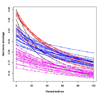
The result is that you get a plot of mechanical advantage against standardised biting positions. And you can compare how mechanical advantages change along the tooth row (see figure to the right). To be clear again, the 0% tooth row position at the left hand side of the plot is the tooth at the back of the tooth row. As expected, that is where the biting efficiency is highest in any taxon studied. Towards the right hand side of the horizontal axis, we get further along the tooth row towards the front of the snout. As usual in lever mechanics, towards the tip of the snout, mechanical advantage gets lower, or less efficient. This is kind of like trying to cut something tough with a long pair of scissors; it is easy to cut/crush through a strong twig at the back of the scissors but really difficult at the tip of the scissors.

It may occur to some that maybe this profile would be consistent across theropod taxa; after all, they all seem to have longish tooth rows with little variation in tooth row morphology. This is true to some extent that the majority of the profile morphology is relatively similar, in that they are all simple parabolic curves. However, there are major differences in the vertical positioning of the curves, or the absolute values of the mechanical advantages (see figure to the left). This graph is from an older version so it's not exactly the same one in the final paper, but the overall pattern is the same; you see a vertical separation in biting profiles, albeit along a continuum (kind of like a smear I guess). Nonetheless, there are prominently unique profiles. For instance the ones in black, Coelophysis and Syntarsus. These profiles criss-cross the entire vertical spectrum from very high mechanical advantages at the back of the tooth row to very low mechanical advantages at the front of the tooth row. Other theropods do not have this extreme combination.

The ones in red are close but not as extreme. These are represented by the two primitive taxa, Herrerasaurus and Plateosaurus. So this is quite likely the ancestral condition in theropods. Dilophosaurus is also showing a similar profile, consistent with this profile being ancestral (although coelophysoids are an exception). Interestingly, some more derived theropods also share similar profiles with these basal taxa; most notably and surprisingly, Carcharodontosaurus. This type of biting is typified by an extremely high mechanical advantage at the back of the tooth row with relatively high to moderate mechanical advantage at the front of the tooth row. What this reflects is that these taxa have their muscle attachments relatively close to a long tooth row, so the overall mechanical advantage is high at the same time the range in mechanical advantage along the length of the tooth row is high as well. In a way the coelophysoid-type profile is an extreme form of this profile.

The rest of the theropods (the blue and pink ones) are almost indistinct from one another in terms of profile (except for a few), but are different in vertical positions, spread out along a continuum. But there is a noticeable gap, hence the different colour designation. This distinction can be a bit arbitrary so I don't make such a distinction in the paper and is discussed in terms of a gradual spectrum; the high-efficiency end of the spectrum towards the top and the low-efficiency (weak/fast biting) end towards the bottom. The high-efficiency function types (blue) have relatively high mechanical advantages along short tooth rows, while conversely the weak/fast function types (pink) have low mechanical advantages along their short tooth rows.

At this point we are still comparing squiggliness of squiggly lines, so in my next post, I shall introduce what I did in my paper in order to make a more meaningful comparison.
Today sees the publication of the new Journal of Vertebrate Paleontology, and with it my paper on the two best-known brachiosaurs and why they’re not congeneric (Taylor 2009).  This of course is why I have been coyly referring to “Brachiosaurus” brancai in the last few months … I couldn’t bear to make the leap straight to saying Giraffatitan, a name that is going to take me a while to get used to.
But before we go lunging into the details, here is my skeletal reconstruction of Brachiosaurus proper, taken from the paper:
Taylor-SVP-Brachiosaurus-fig7-reconstruction-R3-480px
Skeletal reconstruction of Brachiosaurus altithorax, with Homo sapiens and Canis familiaris for scale, from Taylor (2009:fig. 7). White bones represent the elements of the holotype FMNH P 25107. Light grey bones represent material referred to B. altithorax: the Felch Quarry skull USNM 5730, the cervical vertebrae BYU 12866 (C?5) and BYU 12867 (C?10), the "Ultrasauros" scapulocoracoid BYU 9462, the Potter Creek left humerus USNM 21903, left radius and right metacarpal III BYU 4744, and the left metacarpal II OMNH 01138. Dark grey bones modified from Paul's (1988) reconstruction of Giraffatitan brancai. Scale bar equals 2 m.
Those of you familiar with Greg Paul’s classic reconstruction of Giraffatitan brancai will immediately recognise that Real Brachiosaurus is rather differently proportioned, especially in having a longer torso and tail.
This paper has been in the works for some time, and while it was in review and then in press at JVP, it led double life as Chapter 2 of my dissertation.  (For most of its gestation period, the paper’s title was just “Brachiosaurus brancai is not Brachiosaurus“, and the folder where I keep all the project files is still called “bb-is-not-b”).  In the end, I chickened out and went for a longer, more formal, title.
So why are the two species not congeneric?  Well, it’s a long story, and you can read about the detail in the paper, but the bottom line is that virtually every bone that is known from both species differs in significant respects between them.
Of course, I am not the first to suggest that the African brachiosaurid that we know and love isn’t exactly Brachiosaurus.  Credit for that goes to Greg Paul, who more than twenty years ago executed a then-new skeletal reconstruction of that species (the very same reconstruction that is now considered the classic), and in doing so noticed some differences between the American type species Brachiosaurus altithorax and the African referred species “Brachiosaurus” brancai (Paul 1988).  Paul hedged his bets, though: rather than erect a new genus for the African animal, he proposed a subgenus Brachiosaurus (Giraffatitan), so that the full name of the species would become Brachiosaurus (Giraffatitan) brancai; and that of the type species would become Brachiosaurus (Brachiosaurus) altithorax.  Unsurprisingly, this cumbersome nomenclatural scheme did not catch on, and I have not been able to locate a single subsequent reference to these subgenera in the literature.
xxx
Second caudal vertebrae of Brachiosaurus altithorax and Brachiosaurus brancai, equally scaled, from Taylor (2009:fig. 3). A, B, B. altithorax holotype FMNH P 25107; C-G, B. brancai referred specimen HMN Aa. A, C, posterior; B, D, F, right lateral; E, G, anterior. A-B modified from Riggs (1904:pl. LXXV); C-E modified from Janensch (1950a:pl. 2), F-G modified from Janensch (1929:fig. 15). Scale bar equals 50 cm.
That didn’t mean the idea was dead, though: three years later, George Olshevsky’s self-published mega-revision of dinosaur taxonomy proposed raising the name Giraffatitan to genus level (Olshevsky 1991).  Although this genus became popular on the Internet (it cropped up, for example, in Mike Keesey’s much-lamented Dinosauricon web-site), it was almost completely ignored in the technical literature, and even Greg Paul himself subsequently seems to have reverted to using the name Brachiosaurus brancai (e.g. Paul 1994:246).
Why was the new name overlooked?  Partly, I suspect, just because it’s so butt ugly — everyone knows and loves Brachiosaurus brancai, and the name itself has a definite poetry to it that Giraffatitan sorely lacks.  But mostly it’s because Paul didn’t really make a case for the separation that he proposed — wrongly stating, for example, that “the caudals, scapula, coracoid, humerus, ilium, and femur of B. altithorax and B. brancai are very similar” (Paul 1988:7).
That’s how things stood a few years back when I started to take a serious interest in Migeod’s Tendaguru brachiosaurid, which lives in the basement of the Natural History Museum in London.  It quickly started to seem to me that it wasn’t the same thing as what everyone means by Brachiosaurus, but to make sense of it all, I needed first to figure out what the Brachiosaurus actually does mean.  That meant visiting the type material of both species, in Chicago and Berlin, and really looking closely.
Well, I don’t want to go on all day — apart from anything, England play Croatia in a World Cup qualifier in just over an hour — so I’ll just show you some of the the differences between the dorsal vertebrae of the two species.  (You’ll have seen the caudals up above — I just threw them in to break up all that text).
xxx
Dorsal vertebrae of Brachiosaurus altithorax and Brachiosaurus brancai in posterior and lateral views, equally scaled, from Taylor (2009:fig. 1). A, B, E, F, I, J, M, N, B. altithorax holotype FMNH P 25107, modified from Riggs (1904:pl. LXXII); C, D, G, H, K, L, O, P, B. brancai lectotype HMN SII, modified from Janensch (1950a:figs. 53, 54, 56, 60-62, 64) except H, photograph by author. Neural arch and spine of K sheared to correct for distortion. A, D, E, H, I, L, M, P, posterior; B, F, G, J, N, right lateral; C, K, O, left lateral reflected. A, B, dorsal 6; C, D, dorsal 4; E-H, dorsal 8; I-L, dorsal 10; M, N, P, dorsal 12; O, dorsals 11 and 12. Corresponding vertebrae from each specimen are shown together except that dorsal 4 is not known from B. altithorax so dorsal 6, the most anterior known vertebra, is instead shown next to dorsal 4 of B. brancai. Scale bar equals 50 cm.
Lots and lots of differences here — I will quote from the Systematic Paleontology section on the type species: “Postspinal lamina absent from dorsal vertebrae (character 130); distal ends of transverse processes of dorsal vertebrae transition smoothly onto dorsal surfaces of transverse processes (character 142); spinodiapophyseal and spinopostzygapophyseal laminae on middle and posterior dorsal vertebrae contact each other (character 146); posterior dorsal centra subcircular in cross-section (character 151); posterior dorsal neural spines progressively expand mediolaterally through most of their length (“petal” or “paddle” shaped) (character 155); mid-dorsals about one third longer than posterior dorsals (see Paul, 1988:7); middorsals only about 20% taller than posterior dorsals (see Paul, 1988:8); dorsal centra long (Janensch, 1950a:72) so that dorsal column is over twice humerus length (Paul, 1988:8); transverse processes of dorsal vertebrae oriented horizontally (Paul, 1988:8); dorsal neural spines oriented close to vertical in lateral view; dorsal neural spines triangular in lateral view, diminishing smoothly in anteroposterior width from wide base upwards; deep inverted triangular ligament rugosities on anterior and posterior faces of neural spines” …. *gasp*
So anyway: the upshot of all this is that “Brachiosaurus” brancai differs from Brachiosaurus altithorax more than, say, Barosaurus does from Diplodocus; and so it must be placed in its own genus … and that genus has to be Giraffatitan, because of the ICZN’s principle of priority.  And THAT is why the very end of the paper — the last sentence of the Acknowledgements — reads:
Finally, I beg forgiveness from all brachiosaur lovers, that so beautiful an animal as “Brachiosaurus” brancai now has to be known by so inelegant a name as Giraffatitan.
Anyway, go and read the paper; full-resolution figures are freely available if you want to look more closely than the JVP’s PDF allows.

References

(And, yes, Randy, I know what your comment is going to say; go ahead and say it anyway, it’ll give me a chance to explain why your approach is wrong :-))

Fonte: http://svpow.wordpress.com/2009/09/09/brachiosaurus-brancai-is-not-brachiosaurus/

Abelissauro

Abelissauro
 
    O Abelissauro cujo o nome significa " réptil de Abel ", viveu no Brasil e chegava a ter até 3 metros de altura e 7 metros de comprimento e pertencia ao grupo dos dinossauros terópodes Abelisauridae, era um terrível animal carnívoro que caçava por toda a América do Sul, sendo muito comum também na Argentina, o local onde foi encontrado e leva em seu nome cientifico é Comahue.
   O Abelissauro possuía um crânio grande e leve que poderia ultrapassar 80 centímetros de comprimento, possibilitando mover a cabeça com facilidade, isso aliado à pernas musculosas, o transformavam em um temível predador. Ele se alimentava tanto de carne fresca quanto de carniça, .
Dados do Dinossauro:
Nome: Abelissauro
Nome Científico: Abelisaurus comahuensis
Época: Cretáceo
Local onde viveu: América do Sul
Peso: Cerca de 2,5 toneladas
Tamanho: 3 metros de altura e 7 de comprimento
Alimentação: Carnívora

Megalania

Megalania
    
O Megalania cujo nome significa " Grande e Antigo" era um gigantesco lagarto monitor que viveu há aproximadamente 1,6 milhões de anos atrás até 30 mil anos atrás durante o Pleistoceno na Austrália ( Queensland, onde foi descoberto por Owen em 1860 ), era muito parecido com os atuais Dragões de Komodo, apresentando o mesmo modo de vida, alimentação, acasalamento, porém atingia tamanhos muito superiores aos mesmos, chegando a atingir 8 metros da cabeça até a cauda. O Megalania podia caçar ou comer carne putrefata ( carniça ), dos enormes marsupiais que existiam em sua época, além de cobras, outros répteis e pássaros, engolindo até os ossos dos animais e depois disso podendo ficar quase um ano sem precisar se alimentar novamente.
    
Dados do Réptil:
Nome: Megalania
Nome Científico: Megalania prisca
Época: Pleistoceno
Local onde viveu: Austrália
Peso: Cerca de 700 quilos
Tamanho: 8 metros de comprimento
Alimentação: Carnívora


Reino: Animalia
Filo: Chordata
Sub-Filo: Vertebrata
Super-Classe: Tetrapoda
Classe: Reptilia
Sub-Classe: Diapsida
Infra-Classe: Lepidosauria
Ordem: Squamata
Super-Família: Anguinomorphoidea
Família: Varanidae
Xixianykus
    O Xixianykus viveu há aproxmadamente 145 milhões de anos atrás na China, durante o período Cretáceo, media cerca de 0,5 metros de comprimento, um dos menores dinossauros já descobertos e pertencia ao grupo dos Terópodes.
   Era um dinossauro caçador que usava a agilidade para se alimentar de pequenos animais, como insetos, moluscos, anfíbios, répteis, ovos e mamíferos, podendo inclusive escavar, saltar e correr velozmente a procura dos mesmo.
   Seu fóssil, bastante incompleto, foi encontrado na província Chinesa de Henan, a metade superior do animal acabou não sendo preservada, dificultando assim efetuar maiores descobertas. A pesquisa foi patrocinada pela Academia Chinesa de Ciências.
Dados do Dinossauro:
Nome: Xixianykus
Nome Científico: Xixianykus zhangi
Época: Cretáceo
Local onde viveu: Ásia
Peso: Cerca de 10 Kilogramas
Tamanho: 0,5 metros de comprimento
Alimentação: Herbívora
Coahuilaceratops
    O Coahuilaceratops cujo o nome faz referência ao local onde foi encontrado, região do deserto de Coahuila no México, viveu há aproxmadamente 72 milhões de anos atrás na América do Norte durante o período Cretáceo, chegava a medir 2,1 metros de altura e 7 metros de comprimento e pertencia ao grupo dos dinossauros Ceratopsídeos.
   Seus chifres de 1,20 metros de comprimento foram os maiores já encontrados dentre os Ceratopsídeos, os quais provavelmente eram utilizados para defesa contra predadores e para combate entre machos em dispustas por territórios e acasalamento.
   Durante o período me que o Coahuilaceratops viveu nessa região, a mesma era um estuário úmido com uma grande diversdade de vegetação, onde as águas do oceano se mesclavam com as dos rios formando pequenos pântanos, juntamente com Hadrossaurídeos, evidênciados por diversos restos fóseis encontrados juntos, dentre eles diversas conchas e dinossauros fossilizados. As pesquisas foram financiadas pela Universidade de Utah e pela National Geographic Society.
Dados do Dinossauro:
Nome: Coahuilaceratops
Nome Científico: Coahuilaceratops magnacuerna
Época: Cretáceo
Local onde viveu: América do Norte
Peso: Cerca de 5 toneladas
Tamanho: 2,10 metros de altura e 7 metros de comprimento
Alimentação: Herbívora

Ampelosaurus 


How Ampelosaurus may have looked - Anness Publishing / NHMPL

Aegyptosaurus 


How Aegyptosaurus may have looked.
Buy use of this image from our Picture Library.

Aegyptosaurus

Meaning: Egyptian lizard

Pronounced: ee-JIP-toe-SORE-us


How Aegyptosaurus may have looked.

How Aegyptosaurus may have looked

Show taxonomic details


Length: up to 15 m

Diet: herbivorous


Period: Upper Cretaceous

Ages: Cenomanian-Santonian

Time span: 98-93 Ma


Found in: Egypt

Aegyptosaurus scale

List other dinosaurs like this

There may be more details in Wikipedia

quinta-feira, 23 de setembro de 2010

Malária surgiu com gorilas

23/9/2010

Agência FAPESP – Desta vez, o responsável não é o chimpanzé. Sabe-se que os chimpanzés são a fonte do HIV-1, principal causa da Aids, e se suspeitava que também fossem o reservatório de origem do Plasmodium falciparum, parasita que causa a forma mais severa de malária. Mas um novo estudo indica outro primata.

No caso da malária, quem deu origem a essa forma de malária humana é o gorila. A conclusão está em um artigo publicado como destaque de capa na edição desta quinta-feira (23/9) da revista Nature.

Weimin Liu, da Universidade do Alabama, nos Estados Unidos, e colegas de diversos países analisaram cerca de 3 mil amostras de fezes colhidas em diversos locais na África Central.

Foram identificadas infecções por plasmódio em chimpanzés (Pan troglodytes) e gorilas-do-ocidente (Gorilla gorilla), mas não em gorilas-do-oriente (Gorilla beringei) ou em bonobos (Pan paniscus).

Malária surgiu com gorilas

Estudo feito por grupo internacional conclui que parasita que causa malária em humanos atingiu primeiro os gorilas. Estudo é destaque de capa na Nature (divulgação)

O estudo demonstrou que os parasitas de malária do gorila-do-ocidente, a espécie mais comum do gênero Gorilla, são as que mais se aproximam do parasita que atinge o homem.

Os pesquisadores observaram que infecções por plasmódio em primatas são altamente prevalentes, bastante distribuídas e quase sempre resultado de várias espécies de parasitas.

Análises de mais de 1,1 mil sequências genéticas mitocondriais e nucleares de chimpanzés e gorilas revelaram que 99% estavam agrupadas em seis linhagens específicas de hospedeiros, que representam espécies distintas de Plasmodium dentro do subgênero Laverania.

Os pesquisadores descobriram que uma das linhagens, em gorilas-do-ocidente, continha parasitas que eram praticamente idênticos ao Plasmodium falciparum. “Os resultados indicam que o P. falciparum tem origem em gorilas e não em chimpanzés, bonobos ou antigos humanos”, afirmam os pesquisadores no artigo.

O artigo Origin of the human malaria parasite Plasmodium falciparum in gorillas (doi:10.1038/nature09442), de Weimin Liu e outros, pode ser lido por assinantes da Nature em www.nature.com.

quarta-feira, 22 de setembro de 2010

Por dentro das pequenas moléculas

22/9/2010

Por Alex Sander Alcântara

Agência FAPESP – “A biologia é milenar. O homem já fazia biotecnologia quando começou a produzir vinho, por exemplo”, disse Carlos Bloch Jr., pesquisador da Embrapa Recursos Genéticos e Biotecnologia, unidade da Empresa Brasileira de Pesquisa Agropecuária, durante o Workshop sobre Biodiversidade Marinha, realizado este mês na FAPESP.

O pesquisador apresentou no evento técnicas de prospecção de moléculas, com destaque para a espectrometria de massa para análise e síntese de peptídeos.

A pesquisa coordenada por ele pode servir de base na produção de alimentos que sejam mais bem assimilados, além de possibilitar o desenvolvimento de embalagens com propriedades anticongelantes e antimicrobianas.

As fontes de moléculas são as mais variadas. O grupo utiliza informações moleculares provenientes de bancos de dados e projetos genoma que podem ser sintetizados quimicamente e submetidos a testes de atividade in vitro. Os pesquisadores demonstraram que certas moléculas podem mudar de forma com alterações ambientais e, consequentemente, apresentar nova função.

Um exemplo está no leite materno, que tem como molécula principal uma proteína chamada de caseína. Segundo Bloch Jr., durante o processamento enzimático dessa proteína ocorre a liberação de fragmentos proteicos cuja atividade biológica é completamente diferente da molécula original.

“Parece que há dois sistemas enzimáticos de liberação dessas atividades a partir de algo maior: um sistema do próprio organismo (endógeno) e outro que vem de sua associação simbiótica com uma dada microbiota (exógeno)”, explicou.

Segundo ele, as ferramentas de espectrometria de massa, de ressonância magnética nuclear e de cromatografia, entre outras, mudaram a noção de como fazer prospecção de moléculas. “Atualmente, essas técnicas permitem uma resolução sem precedentes a partir de quantidades nanomolares”, disse.

Ao analisar genomas, os pesquisadores perceberam que precisam investigar à luz dessa capacidade de retirar de moléculas maiores informações que estavam codificadas ou “encriptadas”.

“O que propomos é uma prospecção dentro do próprio genoma, ou seja, saber o que tem dentro do genoma que pode ser liberado enzimaticamente e que apresenta outro tipo de atividade inesperada”, disse, salientando que, apesar disso, nem toda proteína pode gerar peptídeos bioativos.

O pesquisador da Embrapa cita como outro exemplo a uva usada na produção de vinho. “Quando se coloca uma levedura para fermentar o suco da uva, em um primeiro momento as eventuais notas enológicas do produto final (o vinho) só puderam ser ‘liberadas’ da matéria-prima (o suco das uvas) a partir de uma ação enzimática exógena, ou seja, do microrganismo utilizado no processo de fermentação”, disse.

O pesquisador acrescenta que notas de caramelo, ou de frutas vermelhas ou de manteiga, por exemplo, surgiram porque foram “desencriptadas” a partir de moléculas mais complexas, com o auxílio de atividades enzimáticas.

Alguns alimentos, segundo Bloch Jr., têm moléculas que não se sabia que estavam presentes. Essas estão guardadas (“encriptadas”) e são liberadas no local certo por atividades enzimáticas, seja pelo próprio organismo que recebe alimento ou por atividade enzimática da microbiota presente no trato digestório dos animais.

“Quando alguém come um pedaço de carne, sabemos que a digestão gera proteínas. Mas saber isso é pouco. O que organismo faz de fato? Esse processo ainda é uma caixa-preta”, afirmou.

Motor da evolução

22/9/2010

Por Fábio de Castro

Agência FAPESP – O trabalho do cientista australiano John Mattick tem contribuído para derrubar paradigmas tradicionais da genética. Segundo o professor da Universidade de Queensland, em Brisbane, a programação genética dos organismos multicelulares foi essencialmente mal compreendida durante os últimos 50 anos.

O equívoco, conta, residia no pressuposto de que a maior parte da informação era codificada em proteínas por meio do RNA, cujo papel seria reduzido à transcrição desses dados.

As pesquisas coordenadas por Mattick, no entanto, ligaram o RNA não-codificador de proteínas à evolução de organismos complexos, à diversidade biológica e à cognição, contribuindo para o desenvolvimento do campo da epigenética – o estudo da parcela de 99% do genoma que não codifica proteínas.


Motor da evolução

John Mattick, da Universidade de Queensland (Austrália), explica como descobertas recentes em genética abalaram paradigmas de cinco décadas e aponta o RNA regulatório como centro da evolução de organismos complexos (foto: divulgação)

O cientista, que já publicou mais de 180 artigos, esteve no Brasil durante o 56º Congresso Brasileiro de Genética, no Guarujá (SP), onde apresentou, no dia 17 de setembro, a conferência “O papel central do RNA regulatório na evolução e no desenvolvimento humano”.

Em entrevista concedida à Agência FAPESP, Mattick destacou que o RNA não-codificador de proteínas – até há pouco tempo conhecido como “DNA lixo” – tem um papel regulatório tão importante que pode ser comparado a um software que controla todo o sistema dos organismos complexos.

Agência FAPESP – O senhor afirma que passamos, nos últimos anos, por uma grande mudança de paradigma, que está transformando completamente a forma como entendemos a genética desde a descoberta da estrutura do DNA. O que mudou na genética?
John Mattick – O que realmente mudou tudo, na minha opinião, foram duas descobertas. A primeira é a surpreendente observação de que o número de genes não é muito diferente entre animais muito simples – um verme do solo, como o Caenorhabditis elegans, por exemplo – e humanos. Todos temos aproximadamente o mesmo número de genes convencionais codificadores de proteínas. E a maior parte desses genes é muito semelhante, tem funções parecidas e codifica o mesmo tipo de proteínas.

Agência FAPESP – Por que essa descoberta foi tão intrigante?
Mattick – Foi uma descoberta chocante, porque, antes dela, o dogma central da biologia molecular dizia que os genes estavam no DNA, eram expressos por algum tempo no código temporário do RNA e, então, eram traduzidos em proteínas, que executavam as tarefas no sistema. Assim, esperava-se que os humanos tivessem muito mais genes e muito mais proteínas do que um verme, por exemplo. Mas isso não ocorre. Temos uma variação maior de proteínas, mas, essencialmente, o mesmo número e o mesmo tipo de genes que esses animais muito simples. No entanto, há uma enorme diferença de complexidade entre um verme – que tem menos de 1 milímetro de comprimento e alguns milhares de células – e um humano, com mais de 100 trilhões de células. Trata-se de um plano celular muito mais complexo, principalmente ao se levar em conta o funcionamento do cérebro. Mas temos o mesmo número de genes, o que indica fortemente que deve haver outro tipo de informação capaz de resultar na construção de algo tão mais complexo.

Agência FAPESP – E qual foi a segunda descoberta?
Mattick – Foi uma segunda surpresa, coerente com a primeira: a maior parte dos genes presentes no genoma humano não codifica proteína alguma. Parecia ser puro entulho genético – foi chamado de “DNA lixo”. Mas o que é necessário para fazer um organismo mais complexo, como o humano, está sendo transmitido pelo RNA, não pela proteína. Antes disso, todo mundo pensava que o RNA era só um intermediário temporário entre o gene e a proteína.

Agência FAPESP – Esse era o dogma central?
Mattick – Sim, o DNA é transcrito pelo RNA e copiado na proteína. Esse é o dogma e ele está certo: o DNA faz RNA e alguns RNAs fazem proteína. Esse é o fluxo de informação. O erro é que se pensava que a maior parte da informação do RNA fluía pela proteína. Mas parece que, nos humanos, apenas uma quantidade muito pequena das informações vai para a proteína. A maior parte vai ao RNA. E, agora, estamos conseguindo evidências de que esse RNA está envolvido em regulações muito mais sofisticadas do sistema. Então, para entender como os humanos são programados, temos que pensar não apenas em termos de proteínas, que são componentes mecânicos do sistema. Por trás, há uma arquitetura altamente sofisticada que permite decidir quais componentes devem ser expressos e as mais diversas funções.

Agência FAPESP – O senhor compara os sistemas regulatórios do RNA a um sistema computacional avançado.
Mattick – Sim. Podemos fazer uma analogia com um sistema sofisticado que é o Boeing 777. Seus componentes mecânicos já eram conhecidos há 50 anos: motores, jatos, aerofólios, bombas hidráulicas e assim por diante. Mas há um mundo de diferenças entre um avião feito em 2010 e outro de 1960, quando os computadores ainda não eram populares e não se usavam fibras ópticas. Os objetos mais sofisticados na nossa sociedade se tornam cada vez mais ricos em informação. E essa informação que está codificada em computadores é transmitida por fibras e fios, que não são precisamente componentes mecânicos, mas sim aparatos de transmissão. Portanto, ir de um objeto simples para outro mais complexo não é apenas uma questão de fazer novos componentes, mas de expandir os sistemas de controle e a arquitetura, no caso dos aviões, para que os componentes funcionem de modos cada vez mais sofisticados.

Agência FAPESP – Estamos começando a vislumbrar que há algo a mais do que os componentes que são observados?
Mattick – Sim. Se você mostrasse uma aeronave moderna a um engenheiro aeronáutico de 1960, ele seria capaz de entender como ela voa, mas não poderia entender como funciona. Ele precisaria ver sobre o solo, separado do avião, as centenas de quilômetros de fibras ópticas que ele usa, para ter uma ideia. Porque, em 1960, a transmissão digital de informação em alta velocidade não era uma realidade. Então, é como se a biologia ou a evolução tivessem descoberto o caminho dos sistemas digitais de controle de comunicação 1 bilhão de anos antes do esperado. Estamos só começando a perceber isso.

Agência FAPESP – O senhor mencionou em sua palestra no Congresso Brasileiro de Genética que o papel de regulação do RNA é particularmente importante no cérebro. Por quê?
Mattick – As enzimas que fazem a edição de RNA – processo que modifica a sequência de nucleotídeos do RNA mensageiro em relação à sequência de DNA que o codifica – expandiram-se muito durante a evolução dos vertebrados, em especial dos mamíferos e primatas. A edição de RNA ocorre em praticamente todos os tecidos, mas é particularmente ativa no cérebro – e é aproximadamente 30 vezes mais intensa no cérebro humano do que no do rato. Talvez a edição de RNA seja a conexão entre o genoma e o ambiente e sua expansão foi criticamente importante para a evolução da plasticidade e dos mecanismos de aprendizagem e memória. A regulação do RNA parece ser central não apenas para o desenvolvimento, mas também para a capacidade de alterar plasticamente a informação genética codificada.

Agência FAPESP – Quando começaram as descobertas que possibilitaram essa quebra de paradigmas?
Mattick – Comecei a publicar sobre isso em 1994, mas foi há cerca de dez anos que essa informação realmente começou a circular. Em parte, devido à descoberta dos micro-RNAs regulatórios, mas principalmente graças ao Projeto Genoma Humano. Porque todo mundo esperava que o homem tivesse mais genes do que um verme. E não foi o caso. Então, começamos a descobrir que o RNA tinha muito mais importância do que se supunha. Essa revolução vem ocorrendo nos últimos dez anos, mas a maior parte das pessoas envolvidas com a biologia molecular ainda está considerando essa releitura. É algo ainda revolucionário, mas que está sendo cada vez mais aceito.

Agência FAPESP – Qual a consequência dessa mudança de rumo? O que muda na ciência?
Mattick – Acho que o que muda realmente é a ideia simplista de como a genética funciona. Ficamos muito mais sofisticados. É um pouco como ter mudado da física newtoniana para a física relativista – embora essa não seja uma analogia muito boa. Mas, de certo modo, é como se o mundo da programação genética ficasse muito mais sofisticado, complexo e diferente do que pensávamos. Ainda trabalhamos com moléculas, com DNA e com proteínas. Mas o RNA é o grande personagem hoje. Todo mundo pensava que ele era só um intermediário temporário entre o “disco rígido” e a proteína. Mas as pessoas vão começar a considerar que o RNA não é apenas um intermediário e entender que ele é o motor computacional da célula e do desenvolvimento. É também o motor computacional do cérebro. Assim, uma vez que entendermos esse princípio, poderemos começar a explorá-lo.

Agência FAPESP – Como estudar uma genética que assume tamanha complexidade?
Mattick – Algumas pessoas dizem que, com essas descobertas, a genética está ficando complicada demais. Acho que nunca vamos conseguir entender um sistema a menos que entendamos sua complexidade, pelo menos no plano conceitual. Assumindo a complexidade dos princípios, poderemos começar a fazer perguntas que vão mais adiante e trabalhar sobre o que está de fato ocorrendo. Então, é realmente mais complicado, mas o primeiro passo é entender que de fato é mais complicado. E o segundo passo é buscar um meio para explorar esse novo espaço.

Agência FAPESP – Isso tudo muda a maneira de fazer perguntas científicas?
Mattick – Não mudam as questões, mas muda a maneira como vamos procurar por respostas. As grandes questões continuam sendo como o desenvolvimento funciona e como o cérebro funciona. As perguntas ainda são as mesmas. Mas acho que agora temos uma plataforma muito mais bem formada para começar a responder essas perguntas. Há todo um mundo cuja existência nem suspeitávamos. Estamos só começando. Levaremos um longo tempo para sair desse ponto, mas o primeiro passo conceitual é muito importante e ele está sendo dado neste exato momento.